Elbow Fracture
Surgical treatment of distal humerus, radial head, coronoid and olecranon fractures
Overview
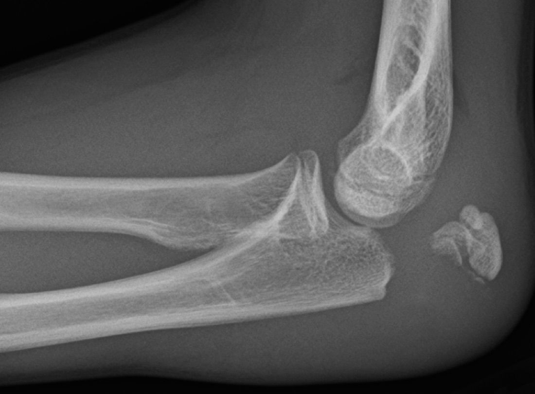
The elbow is a complex joint formed by three bones — the lower end of the upper arm bone (humerus), the inner bone of the forearm (ulna) and the outer bone (radius) — and comprising three separate articulations: the humeroulnar joint (hinge movement: bending and straightening), the humeroradial joint and the proximal radioulnar joint (forearm rotation: supination and pronation). Elbow fractures involve the distal humerus, the olecranon (the upper end of the ulna), the radial head and the coronoid process. They most often result from a fall on an outstretched hand (FOOSH) or a direct blow to the elbow. High-energy injuries (road traffic accidents, falls from height) frequently produce combined fracture–dislocation patterns such as the "terrible triad". Because the elbow is highly prone to stiffness, the fundamental principle of treatment is anatomical reduction with fixation rigid enough to permit early movement.
Anatomy of the Elbow
The projections of the distal humerus (medial epicondyle, lateral epicondyle, trochlea and capitellum) and the olecranon (the prominent bony point at the back of the elbow) provide joint stability. The medial and lateral collateral ligament complexes — particularly the lateral ulnar collateral ligament (LUCL) — form the basis of elbow stability. A healthy elbow allows eating, combing the hair and dressing without pain.
Because the elbow is highly prone to stiffness, the fundamental principle of treatment is anatomical reduction with fixation rigid enough to permit early movement.
Distal Humerus Fractures
These are fractures of the region of the humerus close to the elbow. They usually result from high-energy injuries (in younger patients) or from low-energy falls in older patients with osteoporosis. The AO classification is widely used:
- Supracondylar (extra-articular) fracture: above the joint, proximal to the condyles; particularly common in children
- Transcondylar / intercondylar (T or Y) fracture: involving both the supracondylar region and the joint surface
- Isolated condyle fracture: lateral condyle (the most common, particularly in children) or medial condyle fracture
- Capitellum fracture: the anterior part of the distal humerus that articulates with the radial head
- Medial or lateral epicondyle fracture: usually in adolescents, together with ligament or tendon avulsion
Olecranon Fractures
The olecranon is the prominent bony projection of the ulna at the back of the elbow. It fractures from a direct blow or from sudden contraction of the triceps tendon (avulsion). The Mayo classification is widely used:
- Type I: non-displaced; may be treated non-operatively
- Type II: stable but displaced; surgery is required, with tension band or plate fixation
- Type III: unstable (with associated subluxation or dislocation); plate fixation is standard
- Comminuted fracture: common in older patients with osteoporosis; occasionally excision of the olecranon with reattachment of the triceps tendon, or arthroplasty, may be used
Radial Head Fractures
This is the most common fracture around the elbow. It usually results from a fall on an outstretched hand (FOOSH). The Mason classification is widely used:
- Mason Type I: non-displaced or minimally displaced (<2 mm); treated non-operatively with early movement
- Mason Type II: displaced partial fracture (>2 mm); surgery (screw or mini-plate fixation) is required if it blocks joint movement
- Mason Type III: comminuted, involving the entire radial head; ORIF is used where possible and, rarely, a prosthesis
- Mason Type IV: radial head fracture with elbow dislocation; may be a component of the "terrible triad"
Coronoid Process Fractures
This is a fracture of the small projection on the front of the ulna; it plays a critical role in elbow stability. The Regan–Morrey classification is used:
- Type I: tip fracture only; usually treated non-operatively
- Type II: less than 50% of the coronoid; surgery may be required where there is associated dislocation or instability
- Type III: more than 50% of the coronoid; almost always requires surgery
- Anteromedial fracture: with lateral collateral ligament injury; creates varus instability and requires repair or sometimes reconstruction
Complex Elbow Injuries
These are difficult cases in which more than one structure is injured together. Surgical treatment is required for anatomical reduction and early movement.
- "Terrible triad": elbow dislocation + radial head fracture + coronoid fracture (with lateral collateral ligament rupture)
- Monteggia fracture: proximal ulna fracture + radial head dislocation
- Trans-olecranon fracture–dislocation: an intra-articular olecranon fracture with dislocation of the humeroulnar joint
- Varus posteromedial instability pattern: anteromedial coronoid fracture + LUCL rupture
Classification by Displacement and Joint Involvement
- Non-displaced fracture: the bone fragments have not shifted; some are suitable for non-operative treatment
- Displaced fracture: the fragments have shifted; surgical reduction is required
- Intra-articular fracture: the fracture line extends into the joint surface, increasing the long-term risk of arthritis
- Open fracture: the bone has penetrated the skin; infection risk is high and emergency surgery is required
Symptoms
- Sudden, severe pain: at the moment of injury, prominent around the elbow
- Swelling and bruising: marked bleeding into the joint (haemarthrosis) and oedema within the first hours
- Restricted movement: difficulty bending and straightening the elbow and rotating the forearm
- Deformity: visible in displaced fractures (particularly olecranon and distal humerus fractures)
- Tenderness: pain that increases on palpation of the fracture site
- Crepitus: a grating sensation and sound during movement
- Inability to extend the elbow actively: triceps mechanism insufficiency in olecranon fractures
- Altered sensation or coldness: if there is nerve or vascular injury (particularly the ulnar, median and radial nerves and the brachial artery)
- Open wound: loss of skin integrity in an open fracture
- Loss of joint stability: a feeling of dislocation or of the joint giving way in complex injuries such as the terrible triad
Mechanisms of Injury
- Fall on an outstretched hand (FOOSH): the most common mechanism for radial head and supracondylar fractures
- Direct blow: typical for olecranon fractures (a fall onto the elbow)
- High energy: road traffic accidents, falls from height, motorcycle accidents
- Excessive contraction: sudden triceps contraction (olecranon avulsion fracture)
- Sports injuries: cycling, skiing, motocross, gymnastics
- In children: falls from low height — particularly supracondylar fractures
- Pathological fractures: with minimal trauma in the presence of bone tumour or metastasis
Diagnosis
Diagnosis is based on a detailed history, physical examination and imaging. Examination assesses swelling, haemarthrosis, deformity, points of tenderness, range of motion (flexion–extension and pronation–supination), joint stability (varus/valgus stress tests), neurovascular status (particularly the ulnar nerve — sensation in the little finger and the intrinsic muscles of the hand; the median nerve — thumb flexion; the radial nerve — wrist extension; and the brachial pulse) and skin integrity. Careful monitoring for compartment syndrome is required.
Plain radiographs are the first-line investigation; anteroposterior and lateral elbow views are standard. A "sail sign" (elevation of the anterior fat pad) or a visible posterior fat pad on the lateral view suggests an occult fracture (particularly of the radial head). CT is almost always required for surgical planning in intra-articular fractures (distal humerus, coronoid, comminuted radial head) and complex injuries; three-dimensional CT is critical for assessing fragment displacement. MRI is used to evaluate associated ligament injuries, cartilage damage and occult stress fractures. Angiography or CT angiography may be required where vascular injury is suspected after a high-energy injury.
Non-operative (Conservative) Treatment
Stable, non-displaced fractures that do not disturb the joint surface are suitable for non-operative treatment. Typical candidates are non-displaced Mason Type I radial head fractures, non-displaced olecranon fractures (Mayo Type I) — particularly in lower-demand older patients — non-displaced isolated coronoid tip fractures and some stable distal humerus fractures. Treatment consists of a short period in a splint or cast (10–20 days) followed by graduated active range-of-motion exercises in a purpose-made brace. Prolonged immobilisation markedly increases the risk of elbow stiffness.
Surgical Treatment (ORIF or Arthroplasty)
Open reduction and internal fixation (ORIF) is the standard treatment for displaced or unstable fractures and those that disturb the joint surface. Typical candidates are displaced distal humerus fractures, displaced olecranon fractures, Mason Type II–III radial head fractures (where there is a mechanical block), displaced coronoid fractures, complex elbow injuries (terrible triad, Monteggia, trans-olecranon), open fractures and patients who develop secondary displacement during non-operative treatment. The fundamental aim of treatment is to preserve the patient's own bone and joint surface through anatomical reduction and rigid fixation.
Advantages
The principal advantage is anatomical restoration of the fracture with rigid fixation. This reduces the long-term risk of joint surface incongruity, malunion, non-union and post-traumatic arthritis. Whereas prolonged immobilisation in non-operative treatment can lead to elbow stiffness, surgery allows early movement — which is particularly important in elbow fractures because the elbow is one of the joints most prone to stiffness.
- Anatomical reduction: fragments are restored with millimetre precision and the joint surface is accurately repaired
- Early motion: stable fixation allows elbow range-of-motion exercises to begin within 1–7 days after surgery, markedly reducing the risk of stiffness
- Restoration of stability: ligament injuries and dislocations are repaired, reducing the risk of recurrent dislocation
- Operative time: 60–90 minutes for olecranon and radial head fractures; 120–180 minutes for distal humerus fractures; longer for complex elbow injuries
- Discharge the same day or after 1–2 days (longer in complex cases)
- Elbow function is better preserved in the long term; the risk of post-traumatic arthritis and stiffness is higher after non-operative treatment of displaced intra-articular fractures
Disadvantages
There are surgery-specific risks: wound infection, deep infection, implant failure, nerve injury (particularly the ulnar nerve) and rare vascular injury, together with rare anaesthetic complications.
- Ulnar nerve sensitivity: the ulnar nerve is at risk during distal humerus and olecranon surgery; temporary or permanent numbness may develop. The nerve is protected during surgery or transposed anteriorly.
- Implant irritation: prominence of K-wires and wires beneath the skin at the back of the olecranon is common; implant removal is required in 30–80% of patients. Plates are less irritating.
- Joint stiffness: elbow stiffness may develop despite surgery; additional immobilisation or a late start to movement increases the risk.
- Heterotopic ossification: common in elbow surgery (the risk is three times higher where there is an associated head injury) and may cause loss of movement.
- There is a risk of a poor functional outcome in complex fractures or in patients with significant comorbidity.
Why Surgery?
The elbow is a critical joint for positioning the hand and forearm in space. Even 1–2 mm of joint surface incongruity leads to cartilage wear and post-traumatic arthritis in the long term. Elbow stiffness severely restricts daily activities; the functional range of motion is defined as 30–130° of flexion–extension and 50° of supination to 50° of pronation, and losing this range can prevent a patient from washing their face, combing their hair or eating. In olecranon fractures the integrity of the triceps mechanism is essential for active elbow extension. The surgeon therefore prefers surgical treatment whenever the fracture is displaced and unstable or threatens joint function, and aims for rigid (stable) fixation that permits early movement in order to prevent elbow stiffness.
Who is a Candidate?
Surgery is performed for displaced or unstable elbow fractures and for those that disturb the joint surface.
- Displaced distal humerus fractures
- Mayo Type II–III olecranon fractures
- Mason Type II–III radial head fractures causing a mechanical block or comminuted
- Displaced coronoid fractures (particularly with dislocation)
- Complex elbow injuries (terrible triad, Monteggia, trans-olecranon)
- Open fractures; patients who develop displacement during non-operative treatment
- Patients whose general health is suitable for general anaesthesia and surgery
- Non-operative treatment: stable, non-displaced fractures (Mason Type I radial head, Mayo Type I olecranon, isolated coronoid tip fractures) and selected patients at very high surgical risk
Expected Outcomes
Most patients with a correctly treated elbow fracture achieve a pain-free, functional elbow. Most return to daily activities within 3–6 months; complete recovery may take up to a year in complicated cases. Mild loss of range of motion (particularly 10–20° at the extremes of extension or flexion) is common, but the functional range is preserved in most patients. Recovery may be prolonged after complex elbow injuries and comminuted distal humerus fractures, and a small number of patients are left with persistent stiffness or loss of function. Early surgery, anatomical reduction and disciplined rehabilitation are the principal determinants of success.
Surgery & First 3 Weeks
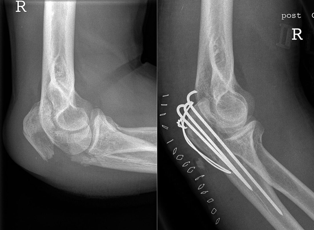
There are two main approaches to the treatment of elbow fractures: non-operative (splint/cast) and surgical (ORIF or arthroplasty). The choice depends on the fracture type, its stability and patient factors. In both approaches it is important to begin movement as early as possible in order to prevent elbow stiffness.
Non-operative Treatment
A splint or cast is used for stable, non-displaced fractures.
- Assessment in the emergency department; reduction under anaesthesia if required
- A short period in a splint or cylinder cast (5–10 days)
- Beginning active range-of-motion exercises as early as possible (within 24–72 hours for radial head fractures in particular)
- Ice, elevation and analgesia
- Follow-up radiographs at weeks 1, 2, 4 and 6 to monitor for displacement
- A forearm sling for as long as needed, usually 1–3 weeks
- Graduated strengthening and physiotherapy
Surgical Treatment — General Information
Surgery is performed under regional (interscalene or supraclavicular) block or general anaesthesia. A regional block is effective for pain control in the first 12–24 hours. Radiographs and CT are reviewed for preoperative planning; three-dimensional CT is used for detailed planning in comminuted distal humerus, coronoid and complex elbow injuries.
Prophylactic antibiotics are given before surgery; routine anticoagulation is not given for upper limb surgery because the risk of DVT is low (except in high-risk patients).
- Duration: 60–90 minutes for olecranon and radial head fractures; 90–120 minutes for distal humerus fractures; 90–120 minutes or more for complex cases such as the terrible triad
- Anaesthesia: regional block or general
- Hospital stay: 1–2 days; longer in complex cases
Surgical Approaches (Incisions)
- Posterior midline approach: standard for distal humerus and olecranon surgery; a longitudinal incision at the back of the elbow
- Olecranon osteotomy: performed to provide wide access to the trochlear joint surface in intra-articular distal humerus fractures; refixed at the end of the procedure with K-wires and a tension band or a plate and screws
- Lateral (Kocher or Kaplan) approach: for the radial head, capitellum and LUCL repair
- Medial approach: for the medial condyle, coronoid and medial collateral ligament repair (protection of the ulnar nerve is required)
- Anterior approach: for distal biceps tendon injury or some coronoid fractures
Distal Humerus Fracture — Technique
- The patient is positioned in the lateral decubitus or supine position (with the elbow free) and a posterior midline incision is made
- An olecranon osteotomy is frequently performed to access intra-articular fractures
- The ulnar nerve is identified and protected, and transposed anteriorly if required
- The joint surface is first reduced anatomically with cannulated screws; the medial and lateral columns are then fixed with parallel or 90-90 (orthogonal) plates (the "dual plating" principle)
- The olecranon osteotomy is refixed with K-wires and a tension band or with a plate
- In exceptional cases in very elderly patients with comminution unsuitable for any form of fixation, total elbow arthroplasty (TEA) may be considered; however, the primary aim in every possible case is to preserve the patient's own anatomy with ORIF
Olecranon Fracture — Technique
- The patient is positioned in the lateral decubitus or supine position and a posterior incision is made
- For simple transverse or short oblique fractures: plate and screw fixation or the tension band technique (two parallel K-wires plus a figure-of-eight wire)
- For comminuted or distally located fractures: an olecranon plate with screws
- For highly comminuted, unreconstructable fractures in older patients: fragment excision with reattachment of the triceps tendon to the ulna
Radial Head Fracture — Technique
- The patient is positioned supine with the forearm pronated; a lateral (Kocher) incision is made
- For Mason Type II (simple displaced) fractures: small screws (Herbert or 1.5–2.0 mm cortical screws) or a mini-plate
- For Mason Type III (comminuted) fractures: reconstruction with ORIF (screws or a mini-plate) is attempted first; a prosthesis is avoided as long as the number and quality of fragments permit reconstruction
- In the rare cases where the radial head cannot be reconstructed, a metallic radial head prosthesis may be considered as a last resort; because the presence of the radial head is critical for joint stability, excision alone is not recommended
Coronoid Process Fracture — Technique
- An anterior or medial approach is used
- Type I (tip) fractures generally do not require surgery
- Type II–III fractures are fixed with sutures or small screws
- Repaired as part of the terrible triad
Terrible Triad — Technique (Elbow Dislocation + Radial Head + Coronoid Fracture)
- A lateral approach is generally used, with an additional medial incision if required
- Sequence: 1) coronoid repair (suture, suture anchor or screw), 2) treatment of the radial head (ORIF or prosthesis), 3) lateral collateral ligament repair, 4) medial collateral ligament repair if required
- Joint stability is tested; an external fixator may be added if instability persists
Monteggia Fracture — Technique (Ulna Fracture + Radial Head Dislocation)
- The ulna is first fixed anatomically with a plate; once the ulna is correctly reduced, the radial head usually relocates spontaneously
- If the radial head does not relocate, open assessment is required for an associated annular ligament tear or soft-tissue interposition
Open Fracture
- Emergency debridement and irrigation, with antibiotic and tetanus prophylaxis
- Depending on the soft-tissue condition, either early definitive fixation or a staged approach with temporary external fixation
Implant Options
- Plates: parallel or 90-90 distal humerus plates (anatomically contoured, locking); olecranon plates; mini-fragment plates for the radial head and coronoid
- Screws: cannulated screws (Herbert, compression); cortical screws (3.5 mm); mini screws (1.5–2.4 mm); locking screws
- K-wires and tension band — the classic technique for olecranon fractures
- Radial head prosthesis (metallic) — a last resort in exceptional cases unsuitable for reconstruction
- Total elbow arthroplasty (TEA) — in highly selected, exceptional cases unsuitable for any form of fixation
- Suture anchors — for coronoid and ligament repair
- External fixator (static or hinged) — for complex unstable fracture–dislocations
Discharge Information
Patients are usually discharged after a hospital stay of 1–2 days; the stay may be longer after complex distal humerus fractures or terrible triad injuries. After surgery the elbow is immobilised in a posterior splint or a hinged elbow brace, with a forearm sling added at the surgeon's discretion.
Before discharge, the physiotherapist teaches how to apply and remove the splint or brace, active shoulder and wrist range-of-motion exercises, use of analgesia and daily transfers. Elbow range-of-motion exercises are usually started within the first 1–7 days as directed by the surgeon; early movement is critical to preventing elbow stiffness.
Ice for 15–20 minutes three to four times a day and elevation of the arm are recommended. Analgesics, anti-inflammatories and gastric protection are prescribed. Routine anticoagulation is not given for upper limb surgery. Dressings are changed regularly; it is important not to wet the wound for the first 10–14 days, and sutures are removed on day 10–14. Supporting the arm on a pillow at night to keep it elevated reduces swelling.
Early Postoperative Period (First 3 Weeks)
The aims during this period are wound healing, control of swelling, pain management, preserving and regaining elbow range of motion, maintaining shoulder and hand movement, and preventing elbow stiffness. Weekly review is recommended.
- Wound monitoring: the skin at the back of the elbow is thin with a limited blood supply. Report discharge, marked redness, increased warmth, wound edge separation or malodour immediately
- Swelling control: elevating the arm above heart level, ice and, if required, a compression glove reduce swelling
- Early motion: with the surgeon's permission, active and active-assisted elbow range-of-motion exercises begin within the first 1–7 days; repeat 4–6 times a day
- Shoulder and wrist movement: active exercises within the splint or brace should be performed three to four times a day; they reduce swelling and prevent stiffness in the adjacent joints
- Pain: once the regional block wears off, prescribed medication, ice and an appropriate resting position control the pain
- Nerve monitoring: the ulnar nerve is the nerve most commonly affected in elbow surgery; numbness in the little and ring fingers and weakness of the intrinsic hand muscles are monitored. Median and radial nerve function are also assessed
- Vascular monitoring: the radial pulse and the colour and temperature of the hand are checked; report coldness, bruising or pallor
Follow-up
- Days 10–14: suture removal and radiographic review
- Weeks 3 and 6: progression of movement and activity
- Month 3: assessment of bone healing
- Month 6 and annually thereafter if required
Risks & Complications
The treatment of elbow fractures is well standardised in modern orthopaedics; however, because the elbow is one of the joints most prone to stiffness, complication rates may be higher than for fractures elsewhere in the limbs. It is important that you understand the possible complications.
Risks Specific to Non-operative (Splint/Cast) Treatment
- Elbow stiffness (contracture): the most common and most important complication; marked loss of range of motion due to prolonged immobilisation
- Inadequate reduction / secondary displacement: fragments shifting within the cast; further surgery may be required
- Skin problems: pressure sores and friction beneath the splint or cast
- Triceps and biceps muscle atrophy
- Sudeck's atrophy (complex regional pain syndrome / CRPS)
- Malunion, non-union and loss of functional range of motion
Risks Specific to Surgery — Common (Under 5%)
- Superficial wound infection (usually treated with antibiotics)
- Postoperative pain, swelling and temporary bruising
- Temporary joint stiffness — resolves with regular physiotherapy
- Transient symptoms related to anaesthesia such as nausea and dizziness
- Temporary numbness of the hand and forearm after a regional block
- Haemarthrosis and prolonged oedema (which may persist for 6–12 months)
Risks Specific to Surgery — Less Common
- Deep infection (osteomyelitis / implant infection): 1–3%; particularly in open fractures; may require revision, implant removal and prolonged antibiotics
- Ulnar nerve injury or late ulnar neuropathy: the ulnar nerve is the nerve most commonly affected in elbow surgery; temporary numbness is common, permanent neuropathy rare
- Median, radial or posterior interosseous nerve injury (with the lateral approach)
- Brachial artery injury (very rare)
- Implant failure: loosening or breakage of screws or plates, particularly with early loading
- Compartment syndrome: rare but very important; increasing pain, tightness and loss of sensation in the forearm; requires emergency fasciotomy
- Heterotopic ossification (bone formation in soft tissue): common in elbow surgery (5–25%), particularly where there is an associated head injury; excision may be required in advanced cases
- Complex regional pain syndrome (CRPS)
- Deep vein thrombosis (DVT) — very rare in the upper limb
- Non-union of an olecranon osteotomy (5–10% in distal humerus surgery)
Long-term Complications
- Elbow stiffness (contracture): the most common long-term complication; marked restriction of movement in 5–20% of patients. Capsular release surgery may be required in advanced cases
- Malunion: mechanical disturbance, pain and an increased risk of arthritis
- Non-union: particularly of the distal humerus and olecranon; increased by smoking and diabetes
- Post-traumatic osteoarthritis: may develop years later after intra-articular fractures
- Chronic instability: a tendency to recurrent dislocation, particularly after complex elbow injuries (terrible triad)
- Chronic pain; late ulnar neuropathy (compression of the ulnar nerve in its groove)
- Loss of strength (particularly grip, gripping and lifting)
Implant-related Problems
- Implant irritation close to the skin: prominence of K-wires and wires beneath the skin at the back of the olecranon; implant removal is required in about 30% of patients (and generally in about 75% after an olecranon tension band)
- Implant migration: displacement of K-wires, which may penetrate the skin
- Plate irritation: a medial distal humerus plate may irritate the ulnar nerve region
- A radial head prosthesis that is too long: disturbance of the joint space and post-traumatic arthritis
- Need for implant removal: usually performed after 9–12 months, once fracture healing is complete
Factors that Increase Risk
- Smoking (markedly increases the risk of wound healing problems, non-union and infection)
- Poorly controlled diabetes (impaired wound healing, risk of infection)
- Osteoporosis (poor implant purchase, secondary displacement)
- Immunosuppressive therapy and rheumatological disease
- Advanced age; high-energy or open fracture
- Associated head injury (increased risk of heterotopic ossification)
- Active or recently treated infection (dental, urinary, skin)
- Prolonged immobilisation (the determining factor for stiffness)
Contact Your Doctor Immediately If You Have
- Sudden increase in pain or swelling
- Fever above 38 °C
- Discharge, malodour or separation of the wound
- Increasing numbness, tingling or weakness in the forearm, hand or fingers
- Bruising, coldness or pallor of the fingers (suspected vascular problem)
- Excessive tightness in the forearm, unbearable pain or pain on straightening the fingers (suspected compartment syndrome)
- Wound edge separation or exposed hardware
- Intense pain or tightness within the splint or cast
Long-term Outcomes
With accurate diagnosis, appropriate treatment and compliant rehabilitation, the great majority of elbow fractures heal without problems. Most patients return to daily activities within 3–6 months, and return to sport is possible within 6–12 months. Complete recovery may take up to a year in some cases; stiffness may persist for 12 months, particularly after complex distal humerus fractures and terrible triad injuries.
There is a risk of post-traumatic arthritis years later after intra-articular fractures; anatomical reduction, early motion and compliant rehabilitation reduce this risk. Stopping smoking, controlling blood glucose and maintaining a healthy weight are critical factors for healing and long-term outcome.
Rehabilitation
Rehabilitation after an elbow fracture is the most important part of recovery. The aims are to control swelling and pain, restore elbow range of motion, regain the strength of the biceps, triceps and forearm muscles, and return the patient to daily life and sporting activity. Because the elbow is one of the joints most prone to stiffness, beginning range-of-motion exercises as soon as it is safe to do so is critical. The functional range of motion is defined as 30–130° of flexion–extension and 50° of supination to 50° of pronation; preserving this range is sufficient for daily activities.
Rehabilitation After Non-operative Treatment
- Days 0–5: Splint or brace. Ice, elevation and pain control. Active shoulder, wrist and finger range-of-motion exercises. Use of a forearm sling.
- Days 5–14: The splint or brace is removed in some fractures (generally earlier in Mason Type I radial head fractures). Active elbow range-of-motion exercises (flexion, extension, supination, pronation) begin, performed 4–6 times a day.
- Weeks 3–6: Range of motion is increased progressively. Active-assisted exercises. The forearm sling is discontinued gradually.
- Weeks 6–12: Full active range of motion is the target. Light strengthening exercises (resistance band, light weights).
- Months 3–6: Advanced strengthening. Full return to daily activities. Return to light recreational sport.
- Months 6–12: Full return to sport-specific drills and contact sport with the surgeon's approval and once functional criteria are met.
Rehabilitation After Surgery (ORIF)
- Days 0–7: Posterior splint or hinged elbow brace. Ice, elevation and pain control. Active shoulder, wrist and finger range-of-motion exercises. With the surgeon's permission, active and active-assisted elbow range-of-motion exercises begin.
- Weeks 1–2: Active elbow range-of-motion exercises 4–6 times a day. Reduction of swelling. Suture removal at days 10–14.
- Weeks 2–6: Range of motion is increased progressively, with full flexion–extension targeted by week 6. The hinged brace is discontinued gradually. Active-assisted exercises.
- Weeks 6–12: Full active range of motion. Light strengthening (resistance band). Pronation–supination exercises intensify. Weight lifting is prohibited.
- Months 3–6: Higher-resistance strengthening. Full return to daily activities. Return to light recreational sport.
- Months 6–12: Full return to sport-specific drills and contact sport with the surgeon's approval. Complete recovery may take 12–18 months after complex elbow injuries.
Special Notes After Complex Elbow Injuries (Terrible Triad)
- If stability has been achieved, elbow range-of-motion exercises begin within the first 1–7 days
- The elbow is exercised from 30° of flexion to 100° using an overhead protocol, with the forearm held in pronation (to protect the lateral collateral ligament)
- Full extension and varus stress are prohibited for the first 6 weeks
- Active strengthening begins at weeks 8–12
Special Notes After Olecranon Fracture
- Active elbow flexion is started early
- Active elbow extension (triceps loading) is restricted until 6–8 weeks to protect the tension band wires
- Strengthening exercises begin at weeks 8–12
Criteria for Return to Sport and Activity
- Full, pain-free elbow range of motion (functional: 30–130° flexion–extension, 50° pronation to 50° supination)
- Forearm and elbow muscle strength at least 85–90% of the opposite arm
- No swelling or pain after activity
- Pain-free completion of sport-specific drills
- Approval of the surgeon and physiotherapist
Recommended Activities
- Walking, stationary cycling, elliptical trainer (low upper limb stress)
- Swimming (once the wound has healed; progressively)
- Low-impact conditioning and strengthening exercise
- Pilates and yoga (with the surgeon's permission, avoiding positions that load the elbow)
- Light hand and forearm strengthening (from month 3)
Activities to Avoid in the First 3–6 Months
- Heavy lifting (particularly above elbow height)
- Exercises that load the elbow such as press-ups and planks
- Throwing sports (baseball, volleyball, handball)
- Tennis and golf (particularly the backhand)
- Contact sport (rugby, handball, basketball)
- Sports with a risk of falling onto the elbow (motocross, mountain biking)
Lifestyle and Recovery Advice
- Perform your home exercise programme every day — early, regular movement is the most effective way to prevent elbow stiffness
- Do not smoke (markedly increases the risk of non-union, wound healing problems and infection)
- Maintain tight blood glucose control if you have diabetes
- Healthy weight: important for overall recovery
- Support the arm on a pillow to keep it elevated during the first weeks (to reduce swelling)
- Do not apply passive force (having someone else forcibly bend or straighten the elbow); this may increase the risk of heterotopic ossification
- Prefer active and active-assisted exercises to static stretching
- Attend your follow-up appointments (weeks 4–6, month 3, month 6, year 1)
Expected Outcomes
Most patients return to pain-free daily activity 3–6 months after treatment of an elbow fracture. Mild loss of range of motion (particularly 10–20° at the extremes of extension or flexion) is common, but the functional range is preserved in most patients.
Return to sport and high-demand activity occurs between months 6 and 12; stiffness and loss of function may persist in the long term after complex elbow injuries (terrible triad, comminuted distal humerus) and high-energy fractures. Patient satisfaction is high with modern treatment methods, early motion protocols and disciplined rehabilitation; mild stiffness is the most common persistent complaint.
Is This Treatment Right for You?
Every patient's condition is different. After a detailed examination and imaging, the treatment plan that suits you best will be prepared.
Would you like more information about Elbow Fracture?
Book Appointment