Cubital Tunnel Syndrome
Treatment of ulnar nerve compression at the elbow causing numbness and hand weakness
Overview
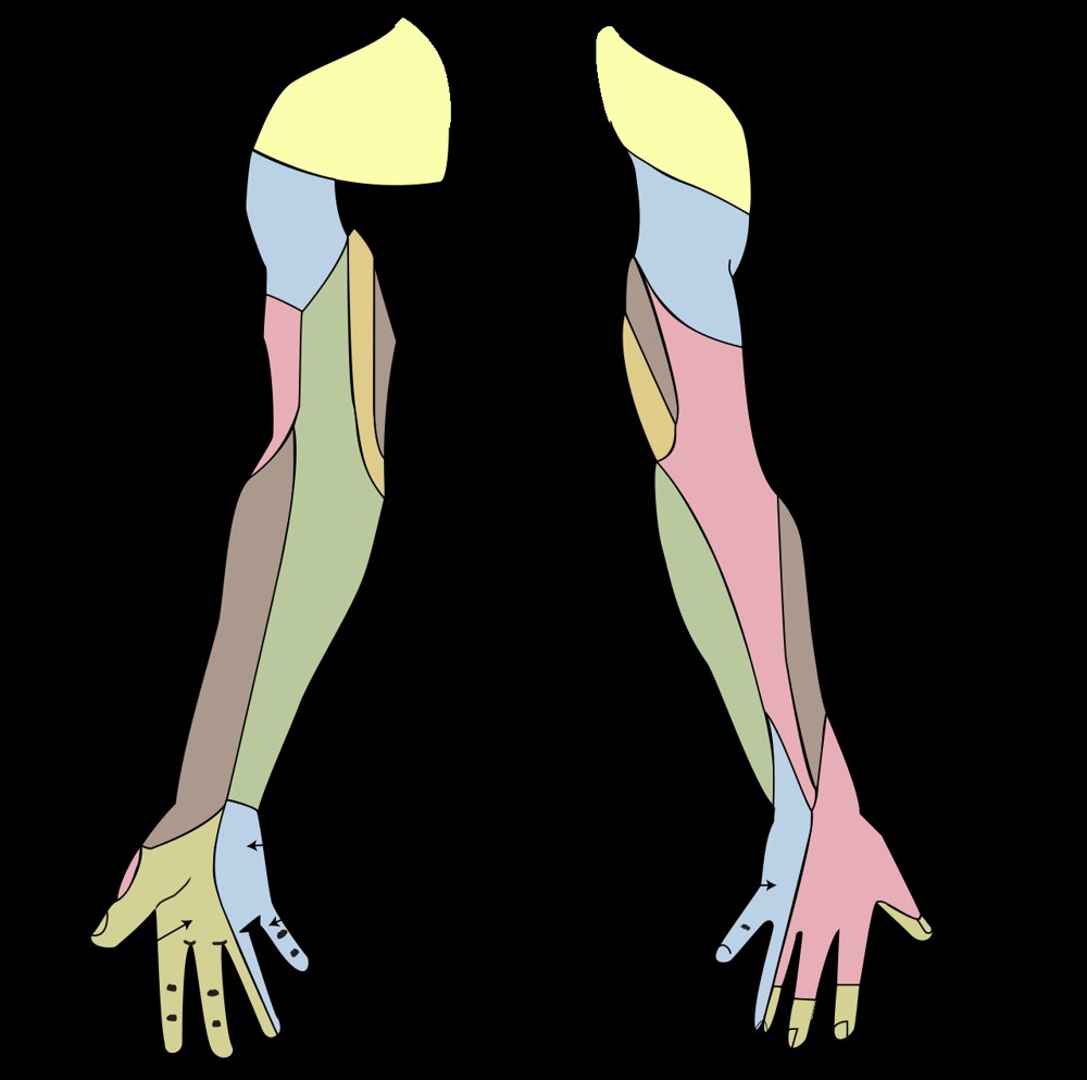
Cubital tunnel syndrome is compression of the ulnar nerve within a narrow canal on the inner side of the elbow, and it is the second most common nerve entrapment of the upper limb after carpal tunnel syndrome. It causes numbness and tingling in the little finger and the inner half of the ring finger and, as it progresses, weakness of the small muscles of the hand. Symptoms characteristically worsen when the elbow is bent, and patients often wake at night with the arm flexed. Early disease settles with simple measures; in neglected cases, permanent wasting of the hand muscles and a claw hand deformity can develop.
Anatomy of the Cubital Tunnel and Mechanism of Compression
The ulnar nerve runs down the inner side of the arm and passes through a groove behind the bony prominence of the inner elbow (medial epicondyle). What produces the electric shock when the elbow is knocked is not bone but the ulnar nerve, lying unprotected beneath the skin.
Beyond the groove the nerve passes under a membranous structure known as Osborne's band, between the two heads of a forearm muscle. The groove and this band together form the cubital tunnel.
When the elbow bends, the volume of the tunnel falls markedly and the nerve is both stretched and compressed. Holding the elbow flexed for long periods — on the telephone, sleeping with the arm bent, resting the elbow while driving — therefore aggravates symptoms directly.
The ulnar nerve supplies sensation to the little finger and the inner half of the ring finger and powers the small muscles within the hand. As compression progresses, grip strength and fine finger control deteriorate.
Risk Factors
- Prolonged elbow flexion: sleeping with the arm bent, long telephone calls, working with the elbow propped on a desk
- Repeated pressure on the inner elbow: leaning on hard surfaces, driving, operating machinery
- Previous elbow fracture or dislocation: distortion of the canal, late (tardy) ulnar nerve palsy
- Elbow arthritis, bone spurs, ganglion cysts within the canal
- Subluxation of the nerve out of its groove on flexion, with repeated friction
- Throwing and racquet athletes: repetitive tension on the inner elbow, sometimes with medial collateral ligament insufficiency
- Diabetes, thyroid disease, rheumatoid arthritis
- Smoking and obesity
Symptoms
- Numbness and tingling in the little finger and inner half of the ring finger: sparing of the thumb and index finger distinguishes it from carpal tunnel syndrome
- Symptoms worse with the elbow bent: on the phone, driving, sleeping
- Waking at night, typical in patients who sleep with the arm flexed
- Pain over the inner elbow radiating along the inner forearm into the hand
- Clumsiness: turning keys, doing up buttons, picking up coins, typing
- Reduced grip and pinch strength, dropping objects
- Wasting of the muscles between the fingers in advanced disease, visible as hollowing on the back of the hand
- Claw hand in advanced disease: the ring and little fingers fail to straighten fully
- Froment's sign: the thumb tip flexes when pinching paper, indicating adductor weakness
Making the Diagnosis
Diagnosis rests on history and examination. Symptoms confined to the ulnar nerve territory and aggravated by elbow flexion are highly indicative.
Examination assesses sensation, finger abduction and adduction strength, and grip. Tingling radiating into the hand when the nerve is tapped at the inner elbow (Tinel's sign) and numbness appearing within a minute of full elbow flexion (elbow flexion test) support the diagnosis. The nerve is palpated during flexion and extension to detect subluxation.
Nerve conduction studies localise and grade the compression and distinguish cervical radiculopathy (C8 - T1), thoracic outlet syndrome and compression in Guyon's canal at the wrist. They are the standard preoperative investigation and may be normal in early disease.
Ultrasound shows thickening of the nerve, subluxation and masses within the canal dynamically. Radiographs demonstrate previous fracture, arthritis and bone spurs.
Non-operative Treatment
In mild and moderate cases without muscle wasting, treatment begins non-operatively. A substantial proportion of patients improve markedly with positional measures alone.
- Night splint or soft elbow wrap holding the elbow at 30 - 45 degrees to prevent excessive flexion during sleep: the most effective single measure
- Activity modification: avoiding leaning on the elbow, using a headset, supporting the arm at the desk
- Elbow pad to protect the nerve from direct pressure during the day
- Nerve gliding exercises, performed gently and without pain
- Anti-inflammatory medication for short-term pain control
- Corticosteroid injection is not routinely recommended: benefit is limited and the superficial nerve is at risk
- Review of technique and training load in athletes
- Control of diabetes and thyroid disease
- Response is assessed after 6 - 12 weeks of consistent treatment
Surgery & First 3 Weeks
Surgery aims to relieve pressure on the ulnar nerve and prevent it being stretched as the elbow moves. The choice of technique depends on whether the nerve subluxates, whether the elbow is structurally abnormal, and whether previous surgery has been performed.
Indications for Surgery
- Inadequate response within three months of consistent non-operative treatment
- Constant numbness and sensory loss
- Moderate to severe compression on nerve conduction studies
- Wasting of the hand muscles or marked loss of grip strength: these should not be delayed
- A structural cause such as a ganglion, bone spur or post-fracture deformity
- Recurrent subluxation of the nerve with elbow flexion
- Surgery before nerve damage is established gives markedly better results
Simple Decompression (in situ release)
Through a 4 - 6 centimetre incision on the inner elbow, Osborne's band and the compressing fascia are divided to open the tunnel. The nerve is left in its own bed.
Because the nerve's blood supply is undisturbed, recovery is quick and rehabilitation short. It is the first choice where the nerve does not subluxate and the elbow is structurally normal; results equal those of transposition with a lower complication rate.
Anterior Transposition
The ulnar nerve is moved out of its groove and placed in front of the medial epicondyle so that it is no longer stretched on flexion. It may be placed under the skin, within the muscle or beneath the muscle.
It is preferred where the nerve subluxates repeatedly, after fracture with deformity, in advanced arthritis and after failed previous surgery.
Because some of the nerve's own vessels must be divided, recovery can be slower, and postoperative pain and stiffness are slightly more likely than after simple decompression.
Medial Epicondylectomy
Part of the bony prominence is removed so that the nerve no longer passes over it. It is used in selected cases and is less favoured today because of the risk of medial collateral ligament injury and elbow instability.
The Operation
- General anaesthesia or a regional block, usually as a day case
- Operating time is 20 - 40 minutes for simple decompression and 45 - 75 minutes for transposition
- A soft dressing is applied; after simple decompression the elbow is usually left free
- A short period of splinting may follow transposition
- Sutures are removed at 12 - 14 days
Risks & Complications
Cubital tunnel surgery is safe, though the complication rate is somewhat higher and recovery longer than for carpal tunnel release. The key issue is that nerve recovery may be incomplete in longstanding cases.
General Surgical Risks
- Wound infection
- Bleeding and haematoma
- Risks related to anaesthesia
- Wound healing problems and scar tenderness
- Temporary elbow stiffness
Risks Specific to This Operation
- Injury to branches of the medial antebrachial cutaneous nerve, causing permanent numbness around the scar or a painful neuroma: the commonest complication
- Persistent or recurrent symptoms: renewed scar formation around the nerve is the commonest cause
- Subluxation of the nerve after simple decompression, sometimes requiring revision
- Renewed compression of the nerve in its new bed after transposition
- Tenderness over the inner elbow when leaning on it
- Permanent muscle wasting and weakness in patients operated late
- Ligament insufficiency and elbow instability after medial epicondylectomy
- Complex regional pain syndrome: rare
Rehabilitation
Rehabilitation aims to restore elbow movement early, prevent scarring around the nerve and rebuild hand strength gradually. The programme differs by technique: movement is free immediately after simple decompression, while the first weeks after transposition are more protected.
0 - 2 Weeks
- The dressing is kept clean and dry and the arm elevated
- Finger, wrist and shoulder movement is free from day one
- After simple decompression the elbow is moved immediately; prolonged immobilisation is avoided
- After transposition the elbow may be splinted briefly, then moved under supervision
- Heavy lifting, pushing and leaning on the elbow are forbidden
2 - 6 Weeks
- Scar massage and moisturising after suture removal
- Restoration of full elbow flexion, extension and forearm rotation
- Ulnar nerve gliding exercises with a therapist, within pain limits
- Return to desk work usually within 1 - 2 weeks
- Leaning on the elbow continues to be avoided
6 - 12 Weeks
- Graded strengthening of grip, finger abduction and the forearm muscles
- Fine motor work: resisted finger spread, therapy putty, key pinch exercises
- Return to heavy manual work and vibrating tools is planned in this period
- Interval throwing programme in throwing and racquet athletes, with medial elbow load increased gradually
Expected Outcomes and the Long Term
- Night waking and positional numbness: markedly reduced within the first weeks
- Intermittent numbness: settles over weeks to months
- Constant sensory loss: nerve recovery is slow and may take 6 - 12 months
- Muscle strength returns largely in patients operated early, and may remain partial where wasting was advanced
- The result depends directly on the degree of nerve damage before surgery
- Recovery is slower and often incomplete in diabetic and longstanding cases
Is This Treatment Right for You?
Every patient's condition is different. After a detailed examination and imaging, the treatment plan that suits you best will be prepared.
Would you like more information about Cubital Tunnel Syndrome?
Book Appointment