Wrist & Hand Fractures
Surgical treatment of distal radius, scaphoid, metacarpal and phalanx fractures
Overview
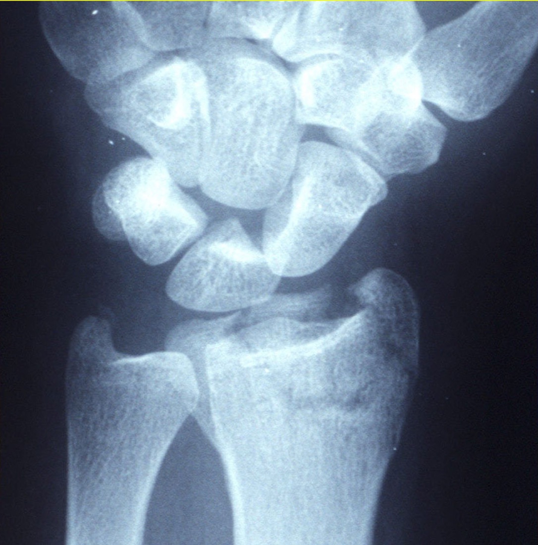
The wrist is a complex joint formed by the two bones of the forearm (radius and ulna) together with the eight carpal bones. The ends of the radius and ulna closest to the wrist (the distal radius and distal ulna) are the most commonly fractured sites. Of the carpal bones, the scaphoid (navicular) is the one most often fractured. The hand contains five metacarpals (palm bones) and fourteen phalanges (finger bones — two in the thumb and three in each of the other fingers). Wrist and hand fractures involve the distal radius, distal ulna, scaphoid, metacarpals and phalanges. They are among the most common orthopaedic injuries and usually result from a fall on an outstretched hand or a direct blow to the hand. Most are suitable for non-operative treatment; however, surgery is required for displaced, unstable or intra-articular fractures.
Anatomy of the Wrist and Hand
A healthy wrist and hand allow gripping, writing, grasping and fine motor tasks — the fundamental movements of daily life — without pain.
Distal Radius Fracture ("Wrist Fracture")
This is the most common fracture in adults. It usually results from a fall on an outstretched hand.
- Colles fracture: dorsally angulated; the most common type (particularly in women over 50)
- Smith fracture: volarly angulated; less common
- Barton fracture: an intra-articular partial fracture–dislocation
- Chauffeur's fracture: a fracture of the radial styloid (the tip)
- Non-displaced / displaced: according to the amount of displacement
- Intra-articular / extra-articular: according to whether the joint surface is involved
Ulna Fracture
This frequently accompanies a distal radius fracture; isolated fractures are uncommon. An ulnar styloid fracture is the most common type and most are treated non-operatively. Fractures of the distal ulna are usually unstable and may require surgery.
Scaphoid (Navicular) Fracture
This is the most commonly fractured carpal bone. Tenderness in the anatomical snuffbox after a fall on an outstretched hand is the characteristic finding. Because the blood supply enters distally and travels proximally, proximal pole fractures carry a high risk of avascular necrosis, and surgery is required if displacement exceeds 2 mm.
- Distal pole fracture: healing is generally good; treated in a cast if there is no displacement
- Waist fracture: the most common type; surgery is required if displacement exceeds 2 mm
- Proximal pole fracture: high risk of non-union and avascular necrosis
Metacarpal Fractures
These are fractures of the palm bones. A punching injury is the most common mechanism.
- Boxer's fracture: a fracture of the neck of the fifth metacarpal (from punching); the most common metacarpal fracture
- Bennett fracture: an intra-articular fracture–dislocation of the base of the first metacarpal
- Rolando fracture: a comminuted intra-articular fracture of the base of the first metacarpal
- Metacarpal shaft or neck fractures
- Metacarpal head fractures (uncommon)
Phalangeal (Finger) Fractures
These are fractures of the finger bones, usually caused by a direct blow or a crush injury.
- Proximal, middle or distal phalangeal fracture
- Mallet fracture: avulsion of the extensor tendon from the dorsal surface of the distal phalanx (the fingertip cannot be straightened)
- Jersey finger: avulsion of the flexor tendon from the volar surface of the distal phalanx
- Intra-articular fractures (condylar and plateau fractures)
- Open fractures (uncommon; may occur with a nail bed injury)
Symptoms
- Pain: at the fracture site, increasing with movement
- Swelling and bruising: oedema developing within the first hours
- Restricted movement: difficulty moving the wrist or fingers
- Deformity: visible in displaced fractures (for example the "dinner fork" deformity of a distal radius fracture)
- Tenderness: pain that increases on palpation of the fracture site
- Weakness of grip: particularly in hand and finger fractures
- Altered sensation: particularly median nerve compression (carpal tunnel-like symptoms in a distal radius fracture)
- Bruising of the nail bed: in distal phalangeal fractures
Mechanisms of Injury
- Fall on an outstretched hand (FOOSH): the most common mechanism for distal radius and scaphoid fractures
- Punching: typical of a boxer's fracture (fifth metacarpal neck)
- Direct blow or crush injury: for phalangeal and metacarpal fractures
- Sports injuries: cycling, skiing, football, volleyball
- Osteoporotic falls: typical of distal radius fractures in older women
Diagnosis
Diagnosis is based on a detailed history, physical examination and imaging. Examination assesses swelling, deformity, points of tenderness, range of motion, grip strength, tendon function (particularly for mallet or jersey finger), neurovascular status (median, ulnar and radial nerves; capillary refill) and skin integrity. Anatomical snuffbox tenderness is specific for a scaphoid fracture.
Plain radiographs are the first-line investigation; anteroposterior, lateral and oblique views are standard. Dedicated scaphoid views are obtained where a scaphoid fracture is suspected; if the initial films are normal they are repeated after 10–14 days, or MRI is used to look for an occult fracture — particularly of the scaphoid. CT is used for surgical planning in intra-articular fractures (especially intra-articular distal radius fractures). MRI is preferred for assessing occult scaphoid fractures, cartilage and soft-tissue damage and tendon injuries.
Non-operative (Conservative) Treatment
Stable, non-displaced fractures — or those brought into an acceptable position by closed reduction — are treated in a cast or splint. Typical candidates are non-displaced distal radius fractures; isolated ulnar styloid fractures; non-displaced scaphoid distal pole or waist fractures; non-displaced or successfully reduced metacarpal neck fractures (a degree of angulation is acceptable in a boxer's fracture in particular); and non-displaced phalangeal fractures. The duration of immobilisation varies by type: 4–6 weeks for the distal radius, 8–12 weeks for the scaphoid and 3–4 weeks for metacarpal and phalangeal fractures.
Surgical Treatment (ORIF / Percutaneous Fixation)
Surgery is the standard treatment for displaced or unstable fractures and for those that disturb the joint surface. Typical candidates are displaced intra-articular distal radius fractures; unstable distal radius fractures (where position cannot be maintained after closed reduction); displaced scaphoid fractures and particularly proximal pole fractures; metacarpal and phalangeal fractures that are displaced or show rotational deformity; Bennett and Rolando fractures; intra-articular phalangeal fractures; and open fractures. The fundamental aims of treatment are anatomical reduction, rigid fixation and early movement.
Advantages
The principal advantage is anatomical restoration of the fracture with rigid fixation. This reduces the risk of malunion, loss of grip strength, rotational deformity of the fingers and post-traumatic arthritis in the long term. Whereas prolonged casting in non-operative treatment can lead to wrist and finger stiffness, surgery allows early movement.
- Anatomical reduction: the joint surface and any extra-articular angulation are corrected with millimetre precision
- Early motion: stable fixation allows wrist or finger range-of-motion exercises to begin within 1–7 days after surgery, reducing the risk of stiffness
- Operative time: 60–90 minutes for a distal radius volar plate; 30–60 minutes for metacarpal or phalangeal fixation; shorter for percutaneous techniques
- Most cases are performed as day cases (discharged the same day)
- Hand function is better preserved in the long term; the risk of post-traumatic arthritis is higher after non-operative treatment of displaced intra-articular distal radius fractures
Disadvantages
There are surgery-specific risks: wound infection, deep infection, implant irritation, rare nerve injury (particularly the median nerve, superficial branches of the radial nerve and the digital nerves) and rare tendon injury, together with rare anaesthetic complications.
- Implant irritation: plates and K-wires placed close to the skin may cause irritation; some patients require hardware removal
- Carpal tunnel syndrome: median nerve compression is common after a distal radius fracture or its surgical treatment (temporary or permanent numbness, night pain)
- Wrist or finger stiffness: may develop despite surgery; additional immobilisation or a late start to movement increases the risk, and intensive physiotherapy may be required
Why Surgery?
The wrist and hand are critical to almost every activity of daily life. Incongruity of 1–2 mm in the distal radius joint surface, or angulation of more than 10°, leads to cartilage wear and post-traumatic arthritis in the long term. In metacarpal and phalangeal fractures, rotational deformity can prevent the fingers from closing properly ("scissoring" — the fingers crossing over one another). Because of their blood supply, scaphoid fractures can lead to non-union and, in the long term, wrist arthritis (SNAC wrist); early surgery reduces the risk of avascular necrosis in displaced or proximal pole fractures. The surgeon therefore prefers surgical treatment where the fracture is displaced and unstable, or where the functional outcome is threatened.
Who is a Candidate?
Surgery is performed for displaced or unstable wrist and hand fractures and for those that disturb the joint surface.
- Displaced intra-articular distal radius fractures; distal radius fractures that are unstable after reduction
- Displaced or proximal pole scaphoid fractures (including non-displaced scaphoid fractures in patients who want early healing and a rapid return to sport)
- Metacarpal and phalangeal fractures with rotational deformity or marked angulation
- Bennett and Rolando fractures; intra-articular phalangeal fractures
- Open fractures
- Mallet or jersey fractures with an associated active tendon injury
- Non-operative treatment: stable, non-displaced fractures, or those that can be brought into an acceptable position by closed reduction
Expected Outcomes
Most patients with a correctly treated wrist or hand fracture achieve a pain-free, functional hand. After a distal radius fracture most return to daily activities within 6–12 weeks, with complete recovery taking up to 6 months. Metacarpal and phalangeal fractures usually heal in 6–8 weeks. Scaphoid fractures take longer to heal (8–16 weeks) and may take longer still for proximal pole fractures. Mild stiffness and a small loss of grip strength may persist, but most patients return to a functional level of use.
Surgery & First 3 Weeks
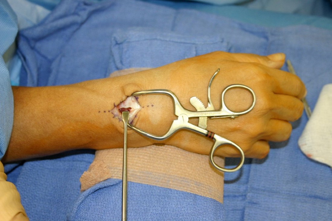
There are two main approaches to the treatment of wrist and hand fractures: non-operative (cast/splint) and surgical (ORIF or percutaneous fixation). The choice depends on the fracture type, its stability and patient factors.
Non-operative Treatment
A cast or splint is used for stable, non-displaced fractures.
- Assessment in the emergency department; closed reduction under haematoma block or regional anaesthesia if required
- Immobilisation: a short arm cast for the distal radius (4–6 weeks); a short or long arm thumb spica cast for the scaphoid (8–12 weeks); an ulnar gutter or finger splint for metacarpal and phalangeal fractures (3–4 weeks)
- Ice, elevation and analgesia
- Follow-up radiographs at weeks 1, 2, 4 and 6 to monitor for displacement
- Graduated movement and physiotherapy once the cast or splint is removed
Surgical Treatment — General Information
Surgery is performed under regional anaesthesia (axillary or supraclavicular block), Bier's block (intravenous regional anaesthesia) or general anaesthesia. Much wrist and hand surgery can be carried out as a day case. Radiographs and, where required, CT or MRI are reviewed for preoperative planning. Prophylactic antibiotics are given; routine anticoagulation is not used for upper limb surgery because the risk of DVT is low.
- Duration: 60–90 minutes for a distal radius volar plate; 30–60 minutes for metacarpal or phalangeal fixation; 30–45 minutes for percutaneous scaphoid screw fixation
- Anaesthesia: regional block or general
- Hospital stay: most patients are discharged the same day; 1 day in complex cases
Distal Radius Fracture — Technique
- Volar (palmar) plate fixation: the most widely used technique. A locking volar plate and screws are applied through a volar approach and the joint surface is reduced anatomically
- Dorsal (back of the wrist) plate: for some dorsally displaced comminuted fractures
- K-wire and cast: percutaneous K-wire fixation after closed reduction in simpler displaced fractures
- External fixator: for open fractures or where there is marked soft-tissue swelling
Distal Ulna / Ulnar Styloid Fracture — Technique
- Most ulnar styloid fractures are treated non-operatively; stability is frequently restored once an associated distal radius fracture is fixed
- Where there is a large, displaced ulnar styloid fracture or instability of the distal radioulnar joint (DRUJ), tension band or small plate fixation is performed
Scaphoid Fracture — Technique
- Percutaneous cannulated screw (Herbert, Acutrak): via a volar or dorsal approach; this has become the standard technique for non-displaced and minimally displaced fractures and shortens the time in a cast
- Open reduction and screw fixation: for markedly displaced, proximal pole or non-united fractures; bone graft is added where required
Metacarpal Fractures — Technique
- Closed reduction and K-wire: percutaneous fixation for most neck and shaft fractures, including boxer's fracture (fifth metacarpal neck)
- Screw fixation: an interfragmentary screw for oblique shaft fractures
- Mini-plates and screws: for comminuted and intra-articular fractures
- Bennett and Rolando fractures: closed reduction with K-wires, or open reduction with a screw or mini-plate
Phalangeal Fractures — Technique
- K-wire or mini-screw: for most displaced shaft or neck fractures
- Mini-plate: for comminuted or intra-articular fractures
- Mallet fracture (with a large dorsal fragment): a screw or K-wire with extension block pinning
- Jersey finger: emergency tendon reinsertion (with a suture anchor, plate and screw or bone tunnel)
Open Fractures
Emergency debridement and irrigation, with antibiotic and tetanus prophylaxis; early fixation where the soft-tissue condition permits.
Implant Options
- Volar or dorsal locking plates — for the distal radius
- Cannulated compression screws (Herbert, Acutrak) — for the scaphoid
- Mini-plates (1.5–2.4 mm) — for metacarpals and phalanges
- K-wires (0.8–1.6 mm) — for percutaneous fixation
- Interfragmentary screws — for oblique fractures
- Suture anchors — for tendon avulsion repair
- External fixator — for open fractures or cases with soft-tissue problems
Discharge Information
Most wrist and hand fracture surgery is performed as a day case and the patient goes home the same day. After surgery the wrist is usually immobilised in a short arm cast, splint or volar splint; an appropriate finger splint is applied for finger fractures. A forearm sling is used to control swelling.
Before discharge the hand therapist provides advice on use of the splint or cast, active shoulder, elbow and finger range-of-motion exercises, elevation of the arm and pain management.
Analgesics, anti-inflammatories and gastric protection are prescribed. Routine anticoagulation is not given for upper limb surgery. Dressings are changed regularly; it is important not to wet the wound for the first 10–14 days, and sutures are removed on day 10–14.
Early Postoperative Period (First 3 Weeks)
The aims during this period are wound healing, control of swelling, pain management, preserving shoulder, elbow and finger movement, and preventing stiffness of the wrist and hand. Weekly review is recommended.
- Wound monitoring: discharge, redness, increased warmth or wound edge separation are monitored for signs of infection
- Swelling control: elevating the arm above heart level, ice and active finger movement reduce swelling
- Movement of the fingers and adjacent joints: active exercises of the free fingers, elbow and shoulder should be performed 4–6 times a day within the splint or cast; this prevents tendon adhesions and stiffness
- Pain: once the regional block wears off, prescribed medication, ice and an appropriate resting position control the pain
- Nerve monitoring: median nerve sensitivity (numbness of the thumb, index and middle fingers, night pain) is common after distal radius surgery. Ulnar and radial nerve function are also assessed
- Vascular monitoring: finger colour, temperature and capillary refill are checked; report coldness, bruising or pallor
Follow-up
- Day 10: suture removal and radiographic review
- Weeks 3–6: cast removal and progression of activity
- Month 3: assessment of bone healing
- Month 6 review if required
Risks & Complications
The treatment of wrist and hand fractures is well standardised in modern orthopaedics and most patients do well. Nevertheless, both cast treatment and surgery carry specific risks.
Risks Specific to Non-operative (Cast/Splint) Treatment
- Inadequate reduction / secondary displacement: fragments shifting within the cast; further surgery may be required
- Pressure sores and friction beneath the cast
- Wrist or finger stiffness: due to prolonged immobilisation
- Loss of grip strength
- Malunion: angulation of the distal radius, or rotational deformity of a metacarpal or phalanx
- Complex regional pain syndrome (CRPS): particularly after distal radius fractures
Risks Specific to Surgery — Common (Under 5%)
- Superficial wound infection (usually treated with antibiotics)
- Postoperative pain, swelling and temporary bruising
- Temporary wrist or finger stiffness — resolves with regular physiotherapy
- Transient symptoms related to anaesthesia such as nausea and dizziness
- Scar discomfort
- Local irritation at K-wire exit sites (with percutaneous techniques)
Risks Specific to Surgery — Less Common
- Deep infection: 1–2%; may require revision and antibiotics
- Median nerve sensitivity / carpal tunnel syndrome: common after distal radius surgery; temporary numbness and night pain
- Injury to the superficial branch of the radial nerve (with dorsal approaches)
- Tendon injury or rupture: particularly rupture of flexor pollicis longus (FPL) after volar plate fixation of the distal radius; irritation of the extensor tendons by long screws
- Complex regional pain syndrome (CRPS)
- Heterotopic ossification (bone formation in soft tissue; rare)
- K-wire migration or breakage
Long-term Complications
- Malunion: ulnocarpal impingement from distal radius angulation; "scissoring" of the fingers from rotational deformity
- Non-union: common in scaphoid fractures in particular (20–30% in displaced and proximal pole fractures); increased by smoking and delayed diagnosis
- Avascular necrosis: death of the bone due to loss of blood supply in scaphoid proximal pole fractures
- Post-traumatic osteoarthritis: may develop years later after intra-articular fractures; SLAC/SNAC wrist is seen particularly with scaphoid non-union
- Persistent wrist or finger stiffness; persistent loss of grip strength
- Chronic pain
- Distal radioulnar joint (DRUJ) instability
Implant-related Problems
- Implant irritation close to the skin: prominence of K-wires beneath the skin; irritation of the tendons by screws
- Need for implant removal: K-wires are usually removed at weeks 3–6 and plates or screws after 6–12 months
- Tendon irritation after a distal radius volar plate (particularly extensor pollicis longus and flexor pollicis longus)
Factors that Increase Risk
- Smoking (the single most important factor for scaphoid non-union)
- Poorly controlled diabetes; osteoporosis
- Immunosuppressive therapy and rheumatological disease
- Advanced age; high-energy or open fracture
- Delayed diagnosis (particularly in scaphoid fractures)
- Active or recently treated infection
- Prolonged immobilisation (the determining factor for stiffness)
Contact Your Doctor Immediately If You Have
- Sudden increase in pain or swelling
- Fever above 38 °C
- Discharge, malodour or separation of the wound
- Increasing numbness, tingling or weakness in the hand or fingers
- Bruising, coldness or pallor of the fingers (suspected vascular problem)
- Excessive tightness or unbearable pain within the cast or splint (suspected compartment syndrome)
- Wound edge separation or exposed hardware
- Severe, burning pain in the fingers (suspected complex regional pain syndrome)
Long-term Outcomes
With accurate diagnosis, appropriate treatment and compliant rehabilitation, the great majority of wrist and hand fractures heal without problems. Most patients return to daily activities within 2–4 months. Mild stiffness and a small loss of grip strength may persist for 6–12 months.
Scaphoid fractures may take longer to heal; delayed diagnosis increases the risk of non-union and of wrist arthritis in the long term. Stopping smoking, early diagnosis and disciplined rehabilitation are critical to long-term outcome.
Rehabilitation
Rehabilitation after a wrist or hand fracture is an integral part of recovery. The aims are to control swelling and pain, restore range of motion, regain grip strength and fine motor skills, and return the patient to daily life and work. Because the wrist and finger joints are highly prone to stiffness, it is critical to move the free joints (shoulder, elbow and the fingers outside the splint) early, even while in a splint or cast. Structured rehabilitation with a hand therapist significantly improves outcomes.
Rehabilitation After a Distal Radius Fracture
- Weeks 0–2: Splint or brace. Ice, elevation and pain control. Active shoulder, elbow and finger range-of-motion exercises 4–6 times a day. After surgery, wound monitoring and suture removal on day 10.
- Weeks 2–6: After surgery, early active wrist range-of-motion exercises begin with the surgeon's permission and the splint is removed in some fractures. In non-operative treatment the cast continues and finger and shoulder exercises are maintained.
- Weeks 6–12: The splint is removed. Active wrist range-of-motion exercises (flexion, extension, supination, pronation, radial and ulnar deviation). Light strengthening; grip exercises (ball squeezing, therapy putty).
- Months 3–6: Higher-resistance strengthening. Full return to daily activities. Return to light recreational sport.
- Months 6–12: Complete functional recovery. Full return to sporting activity with the surgeon's approval.
Rehabilitation After a Scaphoid Fracture
- Weeks 0–6/8: Short or long arm thumb spica cast (the period may be shorter after surgery). Active shoulder, elbow and other finger range-of-motion exercises.
- Weeks 6/8–12: The cast is removed once healing is confirmed radiologically. Active wrist range-of-motion exercises begin.
- Months 3–4: Strengthening and grip exercises. Light recreational activity.
- Months 4–6: Graduated return to sport-specific drills; contact sport with the surgeon's approval.
Rehabilitation After a Metacarpal Fracture
- Weeks 0–3/4: Immobilisation in an ulnar gutter or volar splint. After surgery (K-wire or plate) the splint may be used for a shorter period. Movement of the unaffected fingers continues.
- Weeks 3/4–6: The splint is removed. Active metacarpophalangeal (MCP) and interphalangeal (PIP, DIP) range-of-motion exercises. Light grip exercises.
- Weeks 6–12: Full active range of motion is the target. Graduated strengthening and grip exercises.
- Months 3–6: Complete functional recovery; full return to daily and sporting activity.
Rehabilitation After a Phalangeal Fracture
- Weeks 0–3: Finger splint or buddy taping to the adjacent finger. An appropriate splint after surgery.
- Weeks 3–6: The splint is removed. Active finger range-of-motion exercises; tendon gliding exercises.
- Weeks 6–12: Graduated strengthening. An extension splint may be needed for 6–8 weeks after a mallet fracture.
- Months 3–6: Complete functional recovery; return to all activities.
Criteria for Return to Sport and Activity
- Full, pain-free wrist and finger range of motion
- Grip strength at least 85–90% of the opposite hand
- No swelling or pain after activity
- Radiological confirmation that bone healing is complete (particularly in scaphoid fractures)
- Pain-free completion of sport-specific drills
- Approval of the surgeon and physiotherapist
Recommended Activities
- Walking, stationary cycling, elliptical trainer (low upper limb stress)
- Swimming (once the wound has healed; progressively)
- Low-impact conditioning
- Ball squeezing, therapy putty and finger exercise devices (for functional recovery)
Activities to Avoid in the First 3–6 Months
- Heavy lifting
- Sports with a risk of falling onto the wrist (cycling, skiing, skating)
- Exercises that load the wrist such as press-ups and planks
- Sports involving punching (boxing, martial arts)
- Contact sport
Lifestyle and Recovery Advice
- Perform your home exercise programme every day — early, regular movement is the most effective way to prevent stiffness
- Do not smoke (it markedly increases the risk of non-union, particularly in scaphoid fractures)
- Control your blood glucose if you have diabetes
- Support the arm on a pillow to keep it elevated during the first weeks (to reduce swelling)
- Avoid passive forcing; prefer active exercises
- Attend your follow-up appointments
Expected Outcomes
Most patients return to pain-free daily activity 2–4 months after treatment of a wrist or hand fracture. Mild loss of range of motion and small reductions in grip strength are common, but functional use is preserved in most patients. Complete recovery after a distal radius fracture may take up to 6 months.
With correct treatment and patient rehabilitation, most patients with a scaphoid fracture recover fully, although this may take 6–9 months; there is a risk of long-term complications with delayed diagnosis or proximal pole fractures. Early movement, correct splinting and disciplined rehabilitation are the principal determinants of success.
Is This Treatment Right for You?
Every patient's condition is different. After a detailed examination and imaging, the treatment plan that suits you best will be prepared.
Would you like more information about Wrist & Hand Fractures?
Book Appointment