Carpal Tunnel Syndrome
Operative and non-operative treatment of median nerve compression at the wrist
Overview
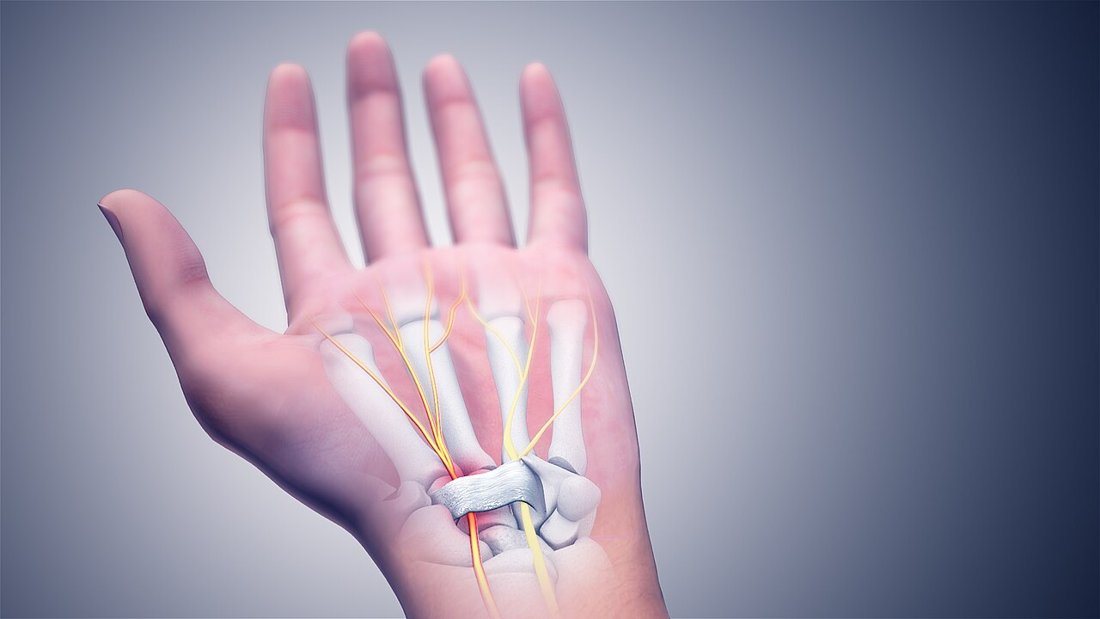
Carpal tunnel syndrome is compression of the median nerve within a narrow canal on the palmar side of the wrist, and it is the most common nerve entrapment of the upper limb. It causes numbness and tingling in the thumb, index and middle fingers, together with burning pain that is typically worse at night. Early symptoms are intermittent and may settle with simple measures; if compression continues, permanent nerve damage, wasting of the thumb muscles and lasting loss of hand strength can develop. Timely diagnosis and treatment at the appropriate stage are therefore decisive.
Anatomy of the Carpal Tunnel and Mechanism of Compression
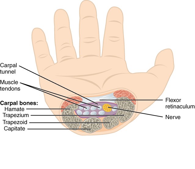
The carpal tunnel is a narrow canal at the level of the wrist whose floor and side walls are formed by the carpal bones and whose roof is the transverse carpal ligament. Nine finger flexor tendons and the median nerve pass through it.
Because its walls are bone and stout ligament, the canal cannot expand. Anything that raises the pressure inside it — swelling of the tendon sheaths, fluid retention, a mass within the canal, or a congenitally narrow canal — presses directly on the median nerve.
Raised pressure first impairs blood flow within the nerve itself; at this stage symptoms are intermittent and vary with hand position. If compression persists, the myelin sheath breaks down and, in advanced disease, nerve fibres are permanently lost and the thenar muscles at the base of the thumb waste away.
The median nerve supplies sensation to the thumb, index finger, middle finger and half of the ring finger, and powers the muscles that lift the thumb out of the plane of the palm and oppose it to the other fingers. The confinement of symptoms to this territory is the basis of the diagnosis.
Risk Factors and Associated Conditions
- Female sex and the 40 - 60 age range: several times more common in women
- Repetitive hand use: prolonged gripping, vibrating tools, work involving constant wrist flexion and extension
- Pregnancy: caused by fluid retention and largely resolves spontaneously after delivery
- Diabetes: increases the nerve's susceptibility to compression, and coexisting polyneuropathy complicates the picture
- Hypothyroidism, rheumatoid arthritis, gout, amyloidosis, chronic kidney failure and dialysis
- Obesity: risk rises markedly with body mass index
- Narrowing of the canal after a wrist fracture or dislocation
- Masses within the canal: ganglion cyst, tenosynovitis, anatomical variants
Symptoms
- Numbness and tingling in the thumb, index and middle fingers: sparing of the little finger is typical and useful in differential diagnosis
- Night pain and waking from sleep: the most characteristic complaint; patients obtain relief by shaking or rubbing the hand
- Symptoms provoked by driving, holding a phone or holding a newspaper
- Burning pain in the hand, which in some patients radiates to the forearm and shoulder
- Reduced grip strength and dropping objects
- Loss of fine dexterity: doing up buttons, threading a needle, picking up coins
- Thenar atrophy in advanced disease: visible wasting at the base of the thumb with permanent loss of strength
- Constant numbness in advanced disease: intermittent symptoms are replaced by persistent sensory loss
Making the Diagnosis
The diagnosis rests primarily on the history and physical examination. The distribution of symptoms, the night-time worsening and the description of relief by shaking the hand are highly indicative.
Examination includes sensory testing, measurement of thumb strength and assessment of the thenar muscle bulk. Provocation tests support the diagnosis: in the Phalen test the wrists are held flexed and numbness appears within a minute; Tinel's sign is tingling radiating into the fingers when the canal is tapped; the carpal compression test reproduces symptoms when pressure is applied over the canal.
Nerve conduction studies (EMG) confirm the diagnosis, grade the severity of compression and distinguish other causes such as cervical radiculopathy or polyneuropathy. They are the standard preoperative investigation in patients scheduled for surgery, although the result can be normal in early disease and does not exclude it.
Ultrasound demonstrates an increased cross-sectional area of the median nerve at the canal entrance and identifies causes within the canal such as a ganglion cyst or tenosynovitis. Plain radiographs are obtained where fracture or arthritis is suspected. Cervical MRI may be required when a disc herniation is in the differential.
Non-operative Treatment
In mild and moderate cases without thenar wasting, treatment begins non-operatively. Success is greatest in patients whose symptoms have lasted less than a year and who have no constant daytime numbness.
- Night splinting: a splint holding the wrist in neutral protects the nerve during sleep, when canal pressure is highest. It is the cornerstone of treatment and should be worn regularly for at least 6 - 8 weeks
- Activity modification: reducing repetitive wrist movements, adjusting work height and keyboard or mouse position, avoiding vibrating tools
- Nerve and tendon gliding exercises: aim to restore free gliding of the median nerve within the canal and are useful in addition to splinting
- Anti-inflammatory medication: provides short-term pain control but does not alter the course of the disease
- Corticosteroid injection into the canal: markedly reduces symptoms and supports the diagnosis, though the effect is temporary in most patients. Repeated injections are avoided
- Treatment of underlying disease: correction of thyroid function, glycaemic control, weight loss
- In pregnancy, splinting and observation are preferred, as the condition usually resolves after delivery
Surgery & First 3 Weeks
Carpal tunnel release divides the transverse carpal ligament completely, widening the canal and relieving pressure on the median nerve. It is one of the most frequently performed and most reliably successful operations in orthopaedics; with the right indication, night pain disappears within the first few days in the great majority of patients.
Indications and Timing
- Inadequate response within 8 - 12 weeks of regular splinting and activity modification
- Constant numbness and sensory loss that persist during the day
- Moderate to severe compression on nerve conduction studies
- Thenar wasting or loss of thumb opposition strength: these call for surgery without delay
- A structural cause such as a mass within the canal or post-fracture narrowing
- Surgery performed before nerve damage is established gives markedly better results than late surgery; once advanced atrophy has developed, pain and numbness may improve while strength does not fully return
Open Carpal Tunnel Release
A 2 - 3 centimetre incision is made in the palm, distal to the wrist crease. The transverse carpal ligament is divided completely along its ulnar border under direct vision, with the median nerve and its motor branch protected.
The main advantage is direct visualisation of both the structure being divided and the nerve being protected. If a ganglion, tenosynovitis or an anatomical variant is found within the canal, it can be addressed at the same sitting. This approach is preferred in revision cases and in patients with a previous wrist fracture.
Endoscopic Carpal Tunnel Release
Through one or two small incisions at the wrist crease, a camera and a purpose-designed blade divide the ligament from inside the canal. Because the skin and deep tissues of the palm are largely spared, scar tenderness is less and return to daily activities may be a few weeks earlier.
At six to twelve months, pain, strength and patient satisfaction are equivalent between the two techniques; the difference lies in early comfort. The endoscopic method works within a narrower field of view, requires experience and is not suitable for every patient.
Anaesthesia and the Operation
- The operation is usually performed as a day case under local anaesthesia or a regional block; general anaesthesia is rarely needed
- Operating time is 15 - 30 minutes
- Patients go home the same day and use the fingers of the operated hand from day one
- When both hands are affected, the more symptomatic side is usually operated first; simultaneous bilateral surgery is possible in selected patients
- Sutures are removed at 10 - 14 days
Risks & Complications
Carpal tunnel release has a low complication rate, with serious complications occurring in well under a few per cent of cases. Nonetheless, the risks should be understood before surgery.
General Surgical Risks
- Wound infection: around 1 per cent and usually superficial
- Bleeding and haematoma
- Anaesthetic risks: very low, as local anaesthesia is used
- Wound healing problems and hypertrophic scar
Risks Specific to This Operation
- Injury to the median nerve or its motor branch: rare, but can cause permanent loss of thumb strength
- Incomplete division of the ligament: the commonest cause of persistent symptoms and may require revision surgery
- Pillar pain: tenderness on either side of the palm, present in a significant minority of patients and usually settling within 3 - 6 months
- Scar tenderness and temporary loss of grip strength: expected during the first 6 - 12 weeks
- Recurrence: uncommon in the long term, usually due to incomplete release or scar formation
- Complex regional pain syndrome: rare, requiring early recognition and intensive hand therapy
- Incomplete resolution of numbness in advanced cases: where nerve damage is established, surgery halts progression but may not reverse the deficit
Rehabilitation
Rehabilitation after carpal tunnel release is straightforward but should not be neglected. The aims are early use of the hand to prevent stiffness and scarring around the nerve, protection of wound healing, and graded recovery of strength.
0 - 2 Weeks
- The dressing is kept clean and dry; the hand is elevated above heart level to reduce swelling
- Finger movement is free and encouraged from day one: full fist and full extension several times a day
- Heavy gripping, pushing and lifting are avoided until the sutures are out
- The wrist is not immobilised in a splint for long periods, as prolonged immobilisation causes stiffness
- Night pain resolves within the first few days in most patients
2 - 6 Weeks
- After suture removal, gentle scar massage and moisturising are started to reduce scar tenderness
- Sensory re-education and tendon gliding exercises continue
- Graded return to daily activities: preparing food, dressing, light housework
- Return to desk work is usually possible within 1 - 2 weeks; manual work takes longer
- Grip strength remains below the preoperative level during this period, which is expected
6 - 12 Weeks
- Graded strengthening: soft ball squeezing, therapy putty, resistance band work for wrist and forearm
- Return to heavy manual work and vibrating tools is usually planned in this period
- Pillar pain, if present, is managed with massage, desensitisation and activity modification
- Grip strength takes an average of three months to reach the preoperative level
Expected Outcomes and the Long Term
- Night pain and waking: usually gone within the first week
- Intermittent numbness: markedly reduced within weeks
- Constant numbness and sensory loss: nerve recovery takes months and may continue for 6 - 12 months
- Thenar atrophy: muscle bulk may partly recover when surgery is early, but can be permanent in advanced cases
- The great majority of patients are satisfied and obtain lasting relief
- Recovery is slower and may be incomplete in patients with diabetes and in long-standing cases
Is This Treatment Right for You?
Every patient's condition is different. After a detailed examination and imaging, the treatment plan that suits you best will be prepared.
Would you like more information about Carpal Tunnel Syndrome?
Book Appointment