Hip Fracture
Surgical treatment of femoral neck, intertrochanteric and subtrochanteric fractures
Overview
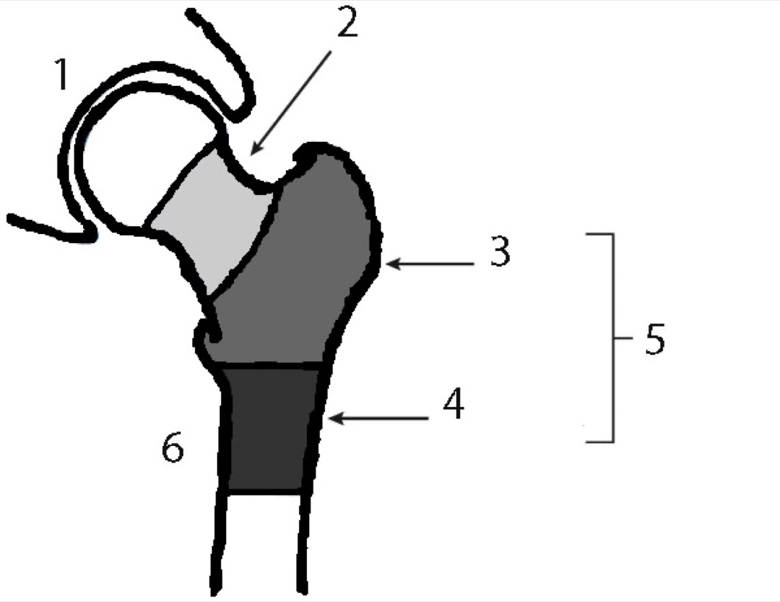
The hip is a ball-and-socket joint formed by the rounded head at the upper end of the thigh bone (femoral head) and the cup-shaped socket in the pelvis (acetabulum). A hip fracture, in the broadest sense, is a fracture of the upper end of the femur (proximal femur), the acetabulum or the pelvic ring. In clinical practice the term "hip fracture" most often refers to a fracture of the upper end of the femur. These fractures are seen particularly in patients over 65 after low-energy falls related to osteoporosis; in younger patients they follow high-energy injuries (road traffic accidents, falls from height). The great majority of hip fractures require surgery; in older patients, early surgery (within the first 24–48 hours) and early mobilisation are of vital importance.
Anatomy of the Hip
The femoral head sits on the femoral neck, which in turn connects to two bony prominences — the greater and lesser trochanters. The blood supply to the femoral head comes largely from vessels that run around the femoral neck; damage to these vessels in a fracture creates the risk of avascular necrosis. The pelvis is a ring-shaped structure formed by the fusion of the iliac, pubic and ischial bones.
A healthy hip allows walking, sitting, bending and climbing stairs — the fundamental movements of daily life — without pain.
Femoral Head and Neck Fractures (Intracapsular)
These lie within the joint capsule. Their most important feature is the high risk of avascular necrosis, because the blood supply to the femoral head comes through this region. The Garden classification is widely used:
- Garden I: non-displaced, valgus impacted (angulated outwards and compressed); the most stable
- Garden II: non-displaced but a complete fracture; the trabecular lines remain intact
- Garden III: partially displaced (with rotation and partial contact)
- Garden IV: completely displaced; the blood supply to the femoral head is very likely disrupted
- Pauwels classification (by fracture angle): Type I (<30°), Type II (30–50°), Type III (>50°); Pauwels III is the most unstable and carries the highest risk of non-union
Intertrochanteric Fractures
These lie outside the joint capsule, in the region between the greater and lesser trochanters. This is the most common type of hip fracture, particularly in older patients. Because the fracture is extracapsular the blood supply to the femoral head is preserved and the risk of avascular necrosis is low. The AO and Evans classifications are widely used:
- Stable (simple, two-part, with an intact posteromedial cortex): treated successfully with a dynamic hip screw (DHS) or an intramedullary nail
- Unstable (comminuted, reverse obliquity, with a posteromedial defect): an intramedullary nail is preferred; the risk of implant failure is higher
- Reverse obliquity fracture: a distinct subgroup requiring an intramedullary nail
Subtrochanteric Fractures
These lie within the 5 cm below the lesser trochanter. They are exposed to high mechanical stress and are biomechanically difficult fractures. Atypical femoral fractures (related to long-term bisphosphonate use) may occur in this region. An intramedullary nail is the gold standard for treatment.
Acetabular Fractures
These are fractures of the hip socket in the side wall of the pelvis. They usually result from high-energy injuries (dashboard injuries in road traffic accidents), although they can also occur after low-energy falls in older patients. The Letournel–Judet classification (10 patterns) is used for surgical planning. Displaced fractures require surgery; acute total hip replacement may be considered in older patients with comminuted fractures.
Pubic and Ischial Ramus Fractures
These are fractures of the slender bony struts at the front and inferomedial part of the pelvic ring (the pubic and ischial rami). They are common in older patients after low-energy falls related to osteoporosis and are generally stable. Most are treated non-operatively: a short period of bed rest, pain control and weight bearing as tolerated are sufficient. In younger patients, where they occur as part of a high-energy pelvic ring injury, surgery (pelvic fixation) may be required, although this is uncommon.
Classification by Displacement and Other Features
- Non-displaced fracture: the bone fragments have not shifted; the great majority still require surgery
- Displaced fracture: the fragments have shifted; surgery is required
- Open fracture: the skin is breached; emergency surgery is required
- Pathological fracture: a fracture occurring with minimal trauma as a result of bone cancer or metastasis
- Atypical femoral fracture: a subtrochanteric or femoral shaft fracture related to long-term bisphosphonate use
- Stress fracture: may be seen in the femoral neck as a result of repetitive loading, particularly in athletes
Symptoms
- Sudden, severe groin or hip pain: after a fall or injury, particularly worse on movement
- Inability to bear weight: unable to stand or walk on the leg
- Deformity: shortening and external rotation of the injured leg (the characteristic finding in a displaced fracture)
- Tenderness: pain that increases on palpation of the groin or greater trochanter
- Restricted movement: difficulty bending or rotating the hip
- Swelling and bruising: oedema developing around the hip (particularly in intertrochanteric fractures)
- Altered sensation or coldness: if there is nerve or vascular injury (uncommon)
- Open wound: loss of skin integrity in an open fracture
- Occult fracture: pain may be mild in some non-displaced femoral neck or pelvic fractures and weight bearing may be preserved; the diagnosis is then made on MRI
Mechanisms of Injury
- Low energy: tripping or falling on level ground (the most common mechanism, particularly in patients over 65 with osteoporosis)
- High energy: road traffic accidents (particularly dashboard injuries — typical for acetabular fractures), falls from height, motorcycle accidents
- Sports injuries (in younger patients)
- Pathological: with minimal trauma in the presence of bone metastasis or tumour
- Atypical: long-term bisphosphonate use (in the subtrochanteric region)
- Stress fracture: in soldiers or athletes who run or march repeatedly
Diagnosis
Diagnosis is based on a detailed history, physical examination and imaging. The mechanism of injury, comorbidities (osteoporosis, diabetes, cardiac and respiratory disease, dementia, smoking) and medication (bisphosphonates, corticosteroids, anticoagulants) are reviewed. Examination assesses leg length discrepancy, external rotation deformity, points of tenderness, range of motion, neurovascular status and skin integrity.
Plain radiographs are the first-line investigation; anteroposterior pelvis, lateral hip and, where required, internally rotated pelvis views are obtained. CT is required for surgical planning in complex fractures and acetabular fractures. MRI is the gold standard where a femoral neck fracture is occult on radiographs, and where a stress fracture or an occult osteoporotic fracture is suspected. A bone scan is an alternative where MRI cannot be performed.
Once a hip fracture is diagnosed, a comprehensive medical assessment (ECG, blood tests, chest radiograph) is performed routinely before surgery. Additional tests are obtained in patients undergoing joint replacement (for example urine culture, or a dental opinion in patients with dental implants).
Non-operative (Conservative) Treatment
Non-operative treatment is used only in selected patients: those at very high surgical risk who are bed-bound with a short life expectancy; some non-displaced impacted or stress fractures; and stable pubic or ischial ramus fractures.
Conservative management consists of bed rest, pain control, compression stockings and anticoagulation, and mobilisation as tolerated. Prolonged bed rest markedly increases the risk of pneumonia, urinary tract infection, pressure sores and deep vein thrombosis, so surgery is preferred in most patients.
Surgical Treatment Options
The great majority of femoral fractures require surgery. The method chosen depends on the fracture type and patient factors: a dynamic hip screw (DHS), an intramedullary hip nail (short or long proximal femoral nail), cannulated screws, hemiarthroplasty (partial hip replacement) or total hip replacement. In acetabular fractures, ORIF or acute total hip replacement may be considered. The great majority of pubic and ischial ramus fractures do not require surgery.
Advantages
The principal advantage is early mobilisation and prevention of the complications of prolonged bed rest (pneumonia, DVT, pressure sores, urinary tract infection, worsening of dementia). In older patients with a hip fracture, early surgery (within the first 24–48 hours) and standing the patient up the day after the operation markedly reduce one-year mortality.
- Anatomical reduction and rigid fixation: the fragments are restored and held stable, making bone healing easier
- Early weight bearing: with modern fixation methods and prostheses, full weight bearing is usually possible the day after surgery
- Lower mortality: the complications of bed rest are minimised
- Pain control: stable fixation markedly reduces pain
- Operative time: 30–60 minutes for cannulated screw fixation; 60–90 minutes for a DHS or intramedullary nail; 60–120 minutes for hemiarthroplasty or total hip replacement
- Hospital stay usually 3–4 days
Disadvantages
- There are surgery-specific risks: wound infection, deep infection, bleeding, anaesthetic complications, deep vein thrombosis and pulmonary embolism. Surgical risks may be higher in older patients because of comorbidities
- Avascular necrosis: may develop even after ORIF, particularly in displaced femoral neck fractures (15–30%), leading to collapse of the femoral head and the need for a prosthesis
- Non-union: 10–30% in femoral neck fractures and lower in intertrochanteric fractures; increased by smoking, diabetes, osteoporosis and inadequate fixation
- Implant failure: screw cut-out (the screw penetrating the femoral head into the joint) and plate loosening may occur in osteoporotic bone
- Acetabular erosion after hemiarthroplasty: movement of the artificial femoral head within the patient's own acetabular cartilage causes wear over time; if pain returns, revision to a total hip replacement may be required
- Risk of dislocation after total hip replacement: 1–5%; the risk is highest in the first 6 weeks and slightly greater with a posterior approach. Hip precautions are required
Why Surgery?
A hip fracture is a life-threatening event, particularly in older patients; one-year mortality is 15–20%, and this rises markedly with prolonged bed rest and is higher still in patients who do not undergo surgery. Early surgery (within the first 24–48 hours) and standing the patient up with full weight bearing the day after the operation reduce complications such as pneumonia, DVT, urinary tract infection, pressure sores and worsening of dementia.
In addition, hip fractures rarely heal well without surgery; malunion, non-union and permanent shortening are common. Surgery is therefore the standard treatment in every patient who is medically fit. In younger patients, early surgical anatomical reduction is critical to preserving the blood supply to the femoral head and restoring normal joint function.
Who is a Candidate?
Surgery is the standard treatment for all displaced femoral fractures, for most non-displaced femoral fractures, and for all patients who are fit for surgery.
- Older patients with osteoporotic femoral neck, intertrochanteric or subtrochanteric fractures
- Younger patients with traumatic femoral fractures
- All patients with an expectation of independent living; those whose comorbidities are under control
- ORIF (cannulated screws) for femoral neck fractures in young, active patients
- Hemiarthroplasty in older, lower-demand patients
- Total hip replacement in active patients with fewer comorbidities and pre-existing hip arthritis
- Non-operative treatment: only in patients at very high surgical risk who are bed-bound; and in stable, non-displaced pubic and ischial ramus fractures
Expected Outcomes
Most patients with a correctly treated hip fracture return to daily activities within 3–6 months. Approximately 75% of older patients return to independent walking; some continue to walk with a stick or frame. Bone healing takes 12–16 weeks in intertrochanteric fractures and 16–24 weeks or longer in femoral neck fractures.
Early weight bearing is possible after hemiarthroplasty and total hip replacement and functional results are generally good. In young, active patients full functional recovery is possible with anatomical reduction; however, if avascular necrosis develops, total hip replacement may be required in the long term.
Surgery & First 3 Weeks
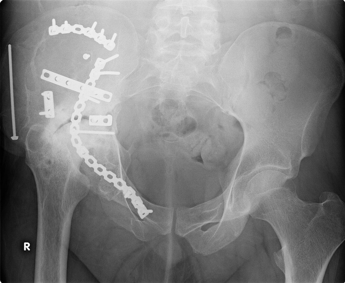
The mainstay of treatment for a hip fracture is surgery. Depending on the fracture type, either fixation (screws, DHS, intramedullary nail) or arthroplasty (hemiarthroplasty, total hip replacement) is chosen. The modern aim is to operate within the first 24–48 hours after the injury.
Non-operative Treatment
Non-operative treatment is used in selected cases: patients at very high surgical risk who are bed-bound with a short life expectancy; some stable non-displaced fractures; and most stable pubic and ischial ramus fractures.
- A short period of bed rest followed by mobilisation as tolerated
- Pain control (analgesics, anti-inflammatories)
- Thromboprophylaxis (anticoagulation, compression stockings)
- Strategies to prevent pressure sores, pneumonia and urinary tract infection
- Physiotherapy: bed exercises, sitting and standing, training with crutches or a frame
- Regular radiographic follow-up (to monitor for secondary displacement)
Surgical Treatment — General Information
Surgery is performed under spinal/epidural or general anaesthesia. The choice is made together with the anaesthetist, taking the patient's comorbidities into account (spinal/epidural may be preferred, particularly in older patients). Radiographs and, where required, CT are reviewed for preoperative planning. Prophylactic antibiotics are given and a plan for thromboprophylaxis is agreed. Surgery is carried out under image intensifier (fluoroscopic) control.
- Duration: 30–60 minutes for cannulated screw fixation; 60–90 minutes for a DHS or intramedullary nail; 60–120 minutes for hemiarthroplasty or total hip replacement
- Anaesthesia: spinal/epidural or general
- Hospital stay: usually 2–5 days; longer in older patients with comorbidities
Surgical Approaches (Incisions)
- Lateral approach: the most common for a DHS and intramedullary nail; a 5–15 cm incision on the outer side of the thigh
- Percutaneous (mini-incision): cannulated screws placed through small incisions
- Posterior approach: widely used for hemiarthroplasty and total hip replacement
- Anterior (Smith–Petersen) approach: a modern choice for total hip replacement; also used for some femoral neck fractures
- Anterolateral (Watson–Jones) approach: an alternative for total hip replacement
Femoral Neck Fracture — Technique
- Young, active patients (generally under 60): closed reduction and percutaneous fixation with cannulated screws. Three 7.0–7.3 mm screws are placed in a triangular or diamond configuration. Urgent surgery may reduce the risk of avascular necrosis
- Older, lower-demand patients with a displaced fracture: hemiarthroplasty (partial hip replacement); a cemented stem is generally preferred
- Active older patients with few comorbidities and pre-existing hip arthritis: total hip replacement; long-term functional results are better than with hemiarthroplasty
- Non-displaced / impacted (Garden I–II): fixation with cannulated screws, a nail or a DHS is successful at any age
Intertrochanteric Fracture — Technique
- For stable fractures, a DHS (dynamic hip screw): a lateral incision is made and a single large lag screw is placed into the femoral head together with a side plate
- For unstable or comminuted fractures, an intramedullary hip nail (PFNA, Gamma nail, TFN type): a short or long nail with distal locking; biomechanically stronger
- For reverse obliquity fractures an intramedullary nail is standard; a DHS is not suitable for these fractures
- In highly comminuted fractures with poor bone quality that are unsuitable for fixation, hemiarthroplasty or a calcar-replacing prosthesis may be used
Subtrochanteric Fracture — Technique
- A long proximal femoral intramedullary nail is the standard treatment; a cephalomedullary nail is used with a lag screw extending into the femoral head
- In atypical femoral fractures, bisphosphonate use is reviewed; prophylactic nailing of the opposite side may be required
- Closed or minimally open reduction; a cerclage wire may be used to hold the reduction if needed
Hemiarthroplasty (Partial Hip Replacement) — Technique
- The patient is positioned in the lateral decubitus or supine position; a posterior or anterior approach is used
- The damaged or fractured femoral head is removed; the acetabular cartilage is left untouched
- The femoral canal is prepared and a femoral stem is inserted (usually cemented in older patients)
- An artificial femoral head is fitted onto the stem; unipolar or bipolar options are available
Total Hip Replacement
The standard total hip replacement technique is used: both the femoral head and the acetabulum are replaced with artificial components. Further detail is given in the 'Hip Replacement' section.
Open Fracture
- Emergency debridement and irrigation, with antibiotic and tetanus prophylaxis
- Depending on the soft-tissue condition, either early definitive fixation or a staged approach with temporary external fixation
Implant Options
- Cannulated screws (7.0–7.3 mm): for femoral neck fractures in young patients
- Dynamic hip screw (DHS) plus side plate: for stable intertrochanteric fractures
- Short intramedullary hip nail (PFNA, Gamma, TFN): for most intertrochanteric fractures
- Long intramedullary nail: for subtrochanteric and reverse obliquity fractures
- Hemiarthroplasty (unipolar or bipolar): in older, lower-demand patients
- Total hip replacement: in active older patients with few comorbidities and pre-existing hip arthritis
- Calcar-replacing prostheses; cerclage wires; external fixator (rarely, in open fractures); trochanteric plate
Discharge Information
Patients are usually discharged after a hospital stay of 2–5 days; the stay may be longer in older patients while their medical condition is stabilised. Discharge is planned to home, to family or to a rehabilitation unit according to the patient's capacity for independent living.
Before discharge, the physiotherapist teaches bed exercises, weight-bearing technique with a frame or crutches (usually full weight bearing with modern fixation and prostheses), transfers, stair technique and the correct technique for daily activities.
After total hip replacement or hemiarthroplasty through a posterior approach, hip precautions apply for the first 6 weeks: do not bend the hip beyond 90°, do not cross the legs, and do not turn the operated leg inwards past the midline. These restrictions are fewer with an anterior approach.
Analgesics, muscle relaxants, gastric protection and anticoagulation (for 4–6 weeks) are prescribed. Compression stockings are worn on the operated leg for 4–6 weeks and on the other leg for 1–2 weeks. Sutures are removed on day 10–14.
Early Postoperative Period (First 3 Weeks)
The aims during this period are wound healing, pain control, maintaining early mobilisation, minimising muscle atrophy, maintaining thromboprophylaxis and closely monitoring general medical condition (particularly in older patients). Weekly review is recommended.
- Wound monitoring: discharge, marked redness, increased warmth or wound edge separation are monitored. It is important not to wet the wound for the first 10–14 days
- Walking and transfers: a frame in the first weeks, then crutches or a stick. Full weight bearing is usually permitted immediately with most surgical methods; partial weight bearing may be required with some comminuted fractures
- Hip precautions: after arthroplasty (particularly a posterior approach), do not bend the hip beyond 90°, cross the legs or turn the operated leg inwards for the first 6 weeks. These restrictions are generally not required after ORIF (nail, screw or plate)
- Pain: prescribed analgesics, ice and an appropriate resting position (lying on your back with a pillow between the legs) control the pain
- Thromboprophylaxis: anticoagulation (4–6 weeks), compression stockings, ankle pumping exercises and early mobilisation
- Exercise: ankle pumping, quadriceps isometric, gluteal isometric and straight leg raise exercises are performed three to four times a day
- Fall prevention: remove hazards such as rugs and cables at home; use a night light, bathroom grab rails and a raised toilet seat
- Medical monitoring: older patients are watched carefully for pneumonia, urinary tract infection, pressure sores and delirium
Follow-up
- Day 7 and days 10–14: suture removal
- Week 6: radiographic review and progression of activity
- Month 3 and month 6 reviews
- Annual follow-up thereafter
Risks & Complications
Surgery for a hip fracture is a life-saving procedure in modern orthopaedics; however, the risk of complications is higher in older patients because of their comorbidities. It is important that you understand the possible complications.
Risks Specific to Non-operative Treatment
- Complications of prolonged bed rest: pneumonia, urinary tract infection, pressure sores, deep vein thrombosis, pulmonary embolism
- Marked muscle atrophy and loss of strength, with difficulty walking after healing
- Increased mortality in bed-bound patients
- Malunion and shortening of the leg
- A marked fall in quality of life and loss of the capacity for independent living
Risks Specific to Surgery — Common (Under 5%)
- Superficial wound infection (usually treated with antibiotics)
- Postoperative pain, swelling and temporary bruising
- Bleeding during surgery; a blood transfusion may occasionally be required
- Temporary restriction of movement or stiffness
- Transient symptoms related to anaesthesia such as nausea, dizziness and urinary retention
- Leg length discrepancy (uncommon and usually able to be compensated for)
- Delirium / confusion (common but temporary in the first days, particularly in older patients)
Risks Specific to Surgery — Less Common
- Deep vein thrombosis (DVT) and pulmonary embolism (PE): among the most important risks of lower limb surgery; largely prevented by anticoagulation, compression stockings and early mobilisation
- Deep infection (periprosthetic infection / osteomyelitis): 1–2%; requires revision, implant removal and prolonged antibiotics
- Nerve injury (particularly the sciatic or femoral nerve; numbness in the lateral femoral cutaneous nerve territory with an anterior approach — very rare)
- Vascular injury (very rare)
- Periprosthetic fracture: a fracture of the femur or pelvis during or after surgery
- Heterotopic ossification (bone formation in soft tissue)
- Serious anaesthetic complications
Complications Specific to Femoral Neck Fractures
- Avascular necrosis (AVN): develops when the blood supply to the femoral head is disrupted; 15–30% after ORIF for displaced fractures and under 10% for non-displaced fractures. If it develops, the femoral head collapses and total hip replacement is required
- Non-union: 10–30% in displaced femoral neck fractures; increased by smoking, diabetes, delayed surgery and inadequate fixation
- Screw cut-out: the screw penetrating the femoral head into the joint in osteoporotic bone
Complications Specific to Intertrochanteric / Subtrochanteric Fractures
- Lag screw cut-out: particularly in osteoporotic bone and with incorrect screw placement
- Femoral shaft fracture: from stress concentration at the tip of an intramedullary nail (very rare)
- Non-union and delayed union in subtrochanteric fractures
- Implant failure (breakage of a plate or screw)
- Leg shortening (particularly in comminuted fractures)
Risks Specific to Hemiarthroplasty
- Acetabular erosion (wear): because the artificial head moves within the patient's own acetabular cartilage, the cartilage and bone may wear over time; if pain returns, revision to a total hip replacement is required
- Dislocation: 1–5%; the risk is highest in the first 6 weeks and slightly greater with a posterior approach
- Long-term functional results are more limited than after total hip replacement
Risks Specific to Total Hip Replacement
- Dislocation: 1–5%; the risk is highest in the first 6 weeks
- Wear: the polyethylene liner may wear over the years
- Loosening of the acetabular or femoral component
Systemic Complications (Particularly Important in Older Patients)
- Pneumonia: related to bed rest and immobilisation
- Urinary tract infection: associated with catheter use
- Pressure sores: during prolonged bed rest
- Delirium / acute confusion: common in the first days after surgery, particularly in older patients and those with dementia
- Cardiac events (heart attack, arrhythmia): in older patients and those with pre-existing heart disease
- Nutritional problems and weight loss
- Loss of independent living (in approximately one third of previously independent patients)
- Mortality: one-year mortality is around 15%; this rises in patients who do not undergo surgery
Factors that Increase Risk
- Advanced age (particularly over 80)
- Smoking (increases the risk of wound healing problems and infection)
- Poorly controlled diabetes; osteoporosis
- Immunosuppressive therapy and rheumatological disease
- Dementia or cognitive impairment (increased risk of delirium)
- Cardiac, respiratory or renal disease; obesity
- Active or recently treated infection (dental, urinary, skin)
- Malnutrition and hypoalbuminaemia
- Delay to surgery (>48 hours) — increases mortality in older patients
Contact Your Doctor Immediately If You Have
- Sudden increase in pain or swelling
- Fever above 38 °C
- Discharge, malodour or separation of the wound
- Firm, one-sided swelling, redness or increased warmth of the leg (suspected DVT)
- Shortness of breath or chest pain (suspected pulmonary embolism)
- Numbness, tingling or loss of strength in the leg
- Sudden deformity of the hip, a feeling of dislocation, or sudden shortening or external rotation of the operated leg
- Acute confusion or hallucinations (suspected delirium)
Long-term Outcomes
With accurate diagnosis, early surgery and compliant rehabilitation, most patients with a hip fracture return to daily activities within 3–6 months. Approximately 75% of older patients return to independent walking; some continue to use a stick or frame.
One-year mortality is 15–20%; this can be reduced by early surgery (within the first 24–48 hours), early mobilisation, good pain control, nutritional support, osteoporosis treatment and fall prevention programmes, but rises in patients who do not undergo surgery. In younger patients, full functional recovery is possible with correct surgery and rehabilitation.
Rehabilitation
Rehabilitation after a hip fracture is the most important part of recovery and is of vital importance, particularly in older patients. The aims are to achieve early mobilisation, control pain, preserve hip range of motion, strengthen the surrounding muscles and return the patient to their previous level of independent living. In the modern approach, standing the patient up the day after surgery is standard.
Rehabilitation After Femoral Fracture Fixation (ORIF)
- Week 0–1: Standing on the first postoperative day; short-distance walking with a frame. Ice and pain control. Ankle pumping, quadriceps and gluteal isometric exercises. The surgeon's weight-bearing protocol is followed.
- Weeks 1–3: Walking distance with the frame is increased progressively. Light strengthening. Graduated return to activities around the house.
- Weeks 3–6: Progression from the frame to crutches and then to a stick. Higher-resistance strengthening; stair practice.
- Weeks 6–12: Most patients discard the stick and walk unaided. Stationary cycling and swimming (once the wound has healed) are recommended.
- Months 3–6: Full return to daily activities. Osteoporosis treatment and fall prevention programmes are continued in older patients.
- Months 6–12: Complete functional recovery. Graduated return to sport in young, active patients with the surgeon's approval.
Rehabilitation After Hemiarthroplasty / Total Hip Replacement
- Week 0–1: Standing on the first postoperative day; short-distance walking with a frame. Full weight bearing is usually permitted immediately. Ankle pumping, quadriceps and gluteal isometric exercises. Hip precautions are applied strictly (after a posterior approach).
- Weeks 1–3: Walking distance with the frame is increased progressively. Graduated return to daily activities. Hip precautions continue.
- Weeks 3–6: Progression from the frame to crutches and then to a stick. Strengthening intensifies. Hip precautions are relaxed gradually.
- Weeks 6–12: Walking with or without a stick. Stationary cycling and swimming are recommended. Hip precautions are mostly lifted.
- Months 3–6: Full return to daily activities. Return to light recreational sport (golf, swimming).
- Months 6–12: Complete functional recovery; most patients begin to use the prosthesis like their own hip.
Hip Precautions — For Patients with a Prosthesis
After hemiarthroplasty or total hip replacement (particularly through a posterior approach), observe the following rules for the first 6 weeks:
- Do not bend the hip beyond 90° (particularly when sitting and bending forwards)
- Do not cross your legs
- Do not turn the operated leg inwards past the midline
- Avoid low chairs, seats and toilets; use raising accessories
- Use long-handled aids for socks, shoes and cutting toenails
- Prefer a shower to a bath; use a shower seat
- Use a pillow between your legs when sleeping
- These restrictions are fewer with an anterior approach; follow your surgeon's instructions
Special Notes on Rehabilitation in Older Patients
- Early mobilisation (the day after surgery) is of vital importance
- Fall prevention: the home environment should be adapted and a stick or frame used correctly
- Osteoporosis treatment should be started (vitamin D, calcium and, where required, a bisphosphonate or denosumab)
- Nutritional support is important; protein intake supports wound healing
- Careful monitoring for dementia and delirium
- Assessment of independent living: some patients may need family support or care services at home
- Rehabilitation unit or home physiotherapy options are considered
Criteria for Return to Sport and Activity (in Young, Active Patients)
- Strength at least 80% of the opposite leg
- Full, pain-free range of motion
- Ability to walk without a walking aid
- Radiological confirmation that bone healing is complete
- No swelling or pain after activity
- Approval of the surgeon and physiotherapist
Recommended Activities
- Walking, light exercise, stationary or outdoor cycling, elliptical trainer
- Swimming (once the wound has healed)
- Light strengthening exercise
- Gardening and housework (excluding heavy lifting)
- Golf (with the surgeon's permission, usually at months 3–6)
Activities Not Recommended or Requiring Caution
- Heavy lifting (particularly from a squat)
- High-impact activities such as jumping (particularly after arthroplasty)
- Tennis, basketball, volleyball (particularly after arthroplasty)
- Contact sport (football, handball, etc.)
- Skiing (particularly at an advanced level)
Lifestyle and Recovery Advice
- Perform your home exercise programme every day — it is the single most important determinant of long-term outcome
- Do not smoke (markedly increases the risk of non-union, wound healing problems and infection)
- Maintain tight blood glucose control if you have diabetes
- Healthy weight: every excess kilogram places additional load on the hip
- Do not neglect osteoporosis treatment (vitamin D, calcium and, where required, a bisphosphonate or denosumab)
- Fall prevention: remove hazards such as rugs and cables at home; use a night light and grab rails in the bathroom
- Treat dental, urinary and skin infections; prophylactic antibiotics may be required if you have a prosthesis
- Attend your follow-up appointments (week 6, month 3, month 6, then annually)
Expected Outcomes
Most patients with a hip fracture return to pain-free daily activity 3–6 months after surgery; complete functional recovery may take 6–12 months. Approximately 75% of older patients return to independent walking, and some continue to use a stick or frame.
Pain control after hemiarthroplasty and total hip replacement is generally good and early return to daily activity is possible. Patient satisfaction is high with both approaches. Preventing falls, treating osteoporosis where present, and attending regular follow-up are the fundamental components of long-term success.
Is This Treatment Right for You?
Every patient's condition is different. After a detailed examination and imaging, the treatment plan that suits you best will be prepared.
Would you like more information about Hip Fracture?
Book Appointment