Rotator Cuff Repair
Arthroscopic repair of the rotator cuff tendons
Overview
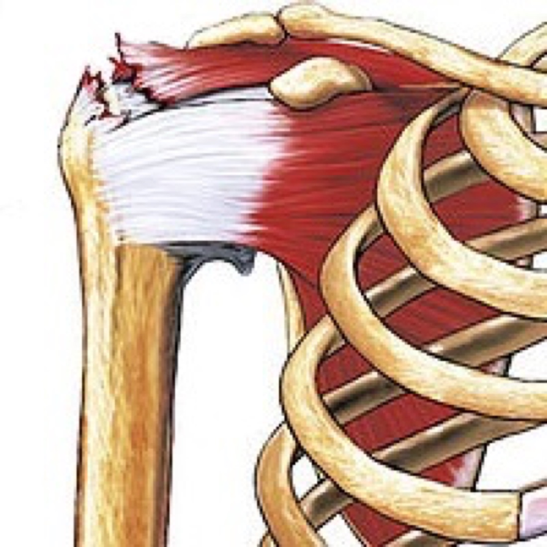
The rotator cuff is a tendon–muscle complex made up of four muscles (supraspinatus, infraspinatus, teres minor and subscapularis) that surrounds the glenohumeral joint. It originates from the shoulder blade (scapula) and inserts onto the head of the upper arm bone (humerus), producing rotational movement of the humerus beneath the acromion.
Function of the Rotator Cuff
- Initiating movement and generating power: the supraspinatus begins elevation of the arm to the side; the infraspinatus and teres minor rotate the arm outwards and the subscapularis inwards.
- Load sharing: working together with the deltoid, it distributes load evenly during overhead activity; if the cuff is weak the deltoid drives the humeral head upwards.
- Joint stability: it keeps the humeral head centred in the glenoid socket, preventing the head from riding out of the socket with every movement of the arm.
- Proprioception: receptors in the tendons and muscles provide a sense of shoulder position, which is disturbed after a tear.
Healing Potential
Blood supply is limited at the site where the tendon attaches to bone (the footprint). Healing depends on the size of the tear, the quality of the tendon and the patient's general health.
- Small tears (<1 cm): blood supply is adequate — very high repair success rate
- Medium tears (1–3 cm): healing is conditional; tendon quality is the determining factor
- Large tears (3–5 cm): higher risk of re-tear
- Massive tears (>5 cm or ≥2 tendons): if tendon quality is inadequate, additional procedures may be required
Types of Tear
- Partial-thickness tear: the tendon is not completely detached from the bone; there is partial damage on the bursal surface, the articular surface or within the tendon substance
- Full-thickness tear: the tear extends through the entire thickness of the tendon; part or all of it is detached from the bone
- Small (<1 cm), medium (1–3 cm), large (3–5 cm), massive (>5 cm or ≥2 tendons)
- Acute (traumatic) tears: occur after a sudden injury
- Degenerative (wear-related) tears: develop over years as a result of age, repetitive overhead activity and impingement syndrome
Symptoms and How the Diagnosis is Made
- Pain over the outer upper shoulder: worse at night and when lying on the affected shoulder
- Difficulty raising the arm: pain and weakness when lifting the arm to the side or overhead
- Difficulty with daily activities: combing hair, reaching high shelves, reaching behind to a back pocket
- Crepitus: crackling or catching during movement
- Acute tear: sudden severe pain and a marked reduction in arm strength
- Silent tear: some degenerative tears may remain asymptomatic for a long time
Diagnosis
Examination includes the Jobe (empty can), drop arm, Hawkins–Kennedy, Neer and lift-off tests; external rotation strength testing assesses the infraspinatus. Plain radiographs show bone architecture, acromial shape and calcific deposits, but do not show the tendon. MRI is the gold standard for assessing the location and size of the tear and the quality of the muscle. Ultrasound provides a rapid, dynamic assessment; its accuracy depends on operator experience. MR arthrography is particularly helpful in partial tears. The definitive decision and treatment plan are made at arthroscopy, with the tear seen directly.
Outcomes are better with early surgery in acute full-thickness tears.
Why Rotator Cuff Repair?
The rotator cuff has critical roles in shoulder stability, initiation of movement, generation of power and control of overhead activity. Preserving as much healthy tendon tissue as possible is therefore of great importance in preventing tear progression, muscle atrophy, fatty infiltration and rotator cuff arthropathy in later years. Studies show that a significant proportion of untreated full-thickness tears enlarge over time, and that the results of delayed repair are poorer. The surgeon therefore prefers to repair the tendon with anchors whenever the tear is amenable to repair.
Advantages
- Most importantly, it prevents tendon retraction, muscle atrophy and shoulder arthritis (rotator cuff arthropathy) over the long term
- Without surgery the tear may enlarge, the tendon may retract and the muscle may undergo fatty infiltration, becoming irreparable
- Operative time: on average 60–90 minutes; general anaesthesia (which may be combined with an interscalene block)
- Hospital stay of 1–2 days
- Small incisions (1 cm), less pain, lower infection risk and a better cosmetic result
Disadvantages
- You will need to wear an abduction sling (a padded sling holding the arm about 30° away from the body) for 6 weeks. Moving the shoulder excessively during this period may prevent healing or cause a re-tear.
- Biological attachment of the tendon to bone takes 3–4 months, so progression to active strengthening is delayed. Shoulder stiffness may develop during this period, but resolves completely with physiotherapy.
- Smoking, diabetes, advanced age and large tear size increase the risk of re-tear; in massive tears this rate may reach 20–30%.
Return to Work and Sport
- Desk-based work: usually week 2 (with the sling)
- Work requiring hand and elbow use: 4 weeks
- Strengthening: month 3
- Graduated return to sport: months 4–6
- Full return to contact, throwing and overhead sport: around month 9
Who is a Candidate?
Arthroscopic rotator cuff repair is performed for tears in which tendon quality is adequate, retraction is limited and there is no significant fatty infiltration of the muscle.
- Patients who have not responded to 6–12 months of non-operative treatment
- Young, active individuals with acute traumatic tears
- Those with marked loss of strength and functional limitation
- People with restricted movement in the dominant arm
- Patients with a large tear but good tendon quality
- Patients whose general health is suitable for general anaesthesia and surgery
Surgery & First 3 Weeks
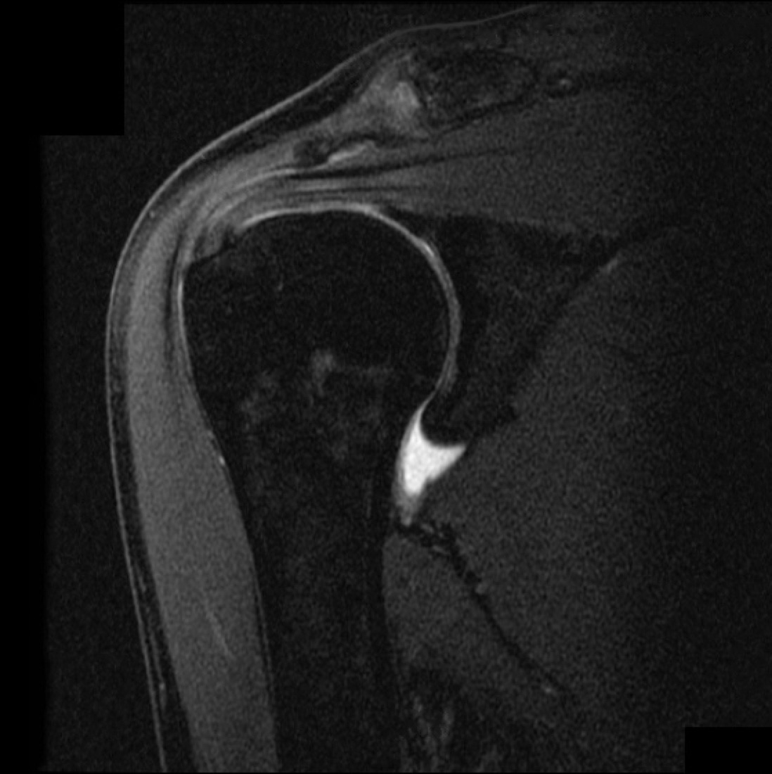
Arthroscopic rotator cuff repair is a procedure in which the tendon that has detached from the bone is first debrided and prepared, then reattached to its original anatomical footprint on the humeral tuberosity with suture anchors and sutures. The shoulder joint and subacromial space are entered arthroscopically through small incisions (approximately 5 mm); the tear is visualised, the space beneath the acromion is decompressed if impingement is present (acromioplasty) and the tendon is repaired with anchors.
General Information About the Operation
- Aim: to preserve the tendon tissue and its continuity as far as possible and restore shoulder function
- Duration: 60–90 minutes, depending on the size of the tear
- Anaesthesia: general (may be combined with an interscalene block)
- Discharge: usually the following day
- Complications: under 5%; most are mild and temporary
Repair Techniques
- Single-row: repair with a single row of anchors placed at the tendon edge. Suitable for small and medium tears.
- Double-row: two rows of anchors, medial and lateral; provides a wider contact area and is preferred for medium to large tears.
- Transosseous-equivalent (suture bridge): a modern technique in which the sutures from the double-row anchors bridge across the repair to distribute compression.
- Acromioplasty: where impingement is present, the undersurface of the acromion is shaved to create space for the tendon.
- Superior capsular reconstruction (SCR): a graft technique used in irreparable massive tears to prevent superior migration of the humeral head.
Discharge Information
Patients are usually discharged after one or two nights in hospital. On the day of surgery you are mobilised with assistance and a sling is fitted. The abduction sling is applied in theatre and should not be removed for approximately 4 weeks; the duration depends on the size of the tear and your surgeon's preference. You may remove the arm from the sling only for washing.
On the first day the physiotherapist teaches elbow, wrist and finger range-of-motion exercises together with scapular isometric and pendulum (Codman) exercises; it is important to perform these 3–4 times a day.
Ice for 15 minutes five times a day is recommended after discharge. Analgesics, muscle relaxants and antibiotic prophylaxis may be prescribed. Dressings should be changed regularly; it is important not to wet the wound for the first 10 days.
Swelling and pain — particularly night pain — are expected in the first 24–48 hours. After the sling is discontinued at week 4, active range-of-motion and light strengthening exercises intensify.
Early Postoperative Period (First 3 Weeks)
You will be discharged wearing the sling, walking, and having learnt your exercises. The operated arm must rest within the sling. Your physiotherapist will show you elbow, wrist and finger range-of-motion exercises together with scapular isometric and pendulum (Codman) exercises; these should be performed five times a day. Weekly review is recommended.
- Wound monitoring: discharge, marked redness and increased warmth are monitored for signs of infection
- Range of motion: being able to bend and straighten the elbow comfortably within the sling and a steady increase in passive range are important. The abduction sling should be worn for 4–6 weeks; active elevation of the arm is prohibited for the first 6 weeks.
- Pain: unless soft-tissue swelling develops around the shoulder, there is a marked reduction within 1–2 weeks. Night pain is the last symptom to settle.
- Nerve monitoring: the axillary and suprascapular nerves are at risk during shoulder surgery. Sensation over the outer upper arm and deltoid strength are monitored; this is rare, but if it occurs, EMG assessment and physiotherapy are required.
Risks & Complications
Arthroscopic rotator cuff repair is generally a safe and effective procedure. It is one of the lower-risk operations in modern shoulder surgery; the rate of serious complications is below 5%.
Common (Under 5%)
- Superficial wound infection (usually treated with antibiotics)
- Postoperative pain, swelling and temporary bruising
- Joint stiffness (a picture resembling frozen shoulder) — resolves with regular physiotherapy
- Temporary weakness — regained with rehabilitation
- Night pain — improves after physiotherapy
Less Common
- Re-tear: 5–10% in small tears and up to 30–40% in massive tears; the risk is higher in older patients, smokers and those with diabetes
- Loosening of a suture anchor
- Persistent shoulder stiffness (frozen shoulder)
- Axillary or suprascapular nerve injury (very rare, <1%)
- Deep infection (very rare)
- Deep vein thrombosis (very rare)
- Chondrolysis — dissolution of the joint cartilage (very rare)
Contact Your Doctor Immediately If You Have
- Sudden increase in pain or swelling
- Fever above 38 °C
- Numbness, tingling or loss of strength in the arm or hand
- Discharge or malodour from the wound
- Sudden marked loss of strength in the arm (suspected re-tear)
- Firm, one-sided swelling and redness of the leg, or shortness of breath
Rehabilitation
Rehabilitation after rotator cuff repair aims to restore range of motion and strength progressively while protecting the biological attachment of the tendon to bone. The programme is advanced according to the size of the tear and the surgeon's protocol.
Weeks 0–4
- Padded sling full time
- Ice, elevation and pain control
- Pendulum (Codman) exercises; elbow, wrist and finger range-of-motion exercises; scapular isometric exercises
- Active elevation of the arm is prohibited; assisted elevation using the other arm or a stick is recommended
Weeks 4–12
- Weeks 4–6: The sling is discontinued. Passive and active-assisted range-of-motion work.
- Weeks 6–12: Progressive increase in flexion and abduction. Active elevation of the arm begins gradually.
Months 3–12
- Months 3–4: Target is full active range of motion. Light rotator cuff and scapular stabilisation strengthening (resistance band). Swimming (progressive) and cycling are recommended. Weight lifting is prohibited.
- Months 4–6: Proprioception and dynamic stabilisation training. Higher-resistance strengthening. Introduction of plyometric exercises.
- Months 6–9: Sport-specific drills; throwing mechanics and overhead movements are introduced gradually. Return to low-contact sport.
- Months 9–12: Full return to contact, throwing and overhead sport with the surgeon's approval. Recovery may take longer after massive tears.
Criteria for Return to Sport
- Rotator cuff and scapular muscle strength at 90% of the opposite shoulder
- Full, pain-free active range of motion
- No painful arc or sensation of impingement
- Pain-free completion of sport-specific drills
- No swelling or pain after activity
Shoulder Health After Repair
A successful rotator cuff repair preserves the tendon and supports long-term shoulder health. The following measures reduce the risk of re-tear and rotator cuff arthropathy:
- Regular rotator cuff and scapular stabilisation exercises
- Correct lifting technique; care when working with weights overhead
- Lower-risk sports: swimming (progressive), cycling, running, elliptical trainer
- Take breaks and stretch during activities requiring repetitive overhead strain
- Stopping smoking (it significantly impairs tendon healing)
- Tight blood glucose control in patients with diabetes
Lifestyle Advice
- Warm up before exercise and stretch afterwards
- Avoid lifting weights overhead for the first 6 months
- Avoid lying on the operated shoulder for the first 3 months; lie on your back or on the opposite side
- Avoid sudden pulling, tugging and movements such as turning a stiff door handle
- Attend your follow-up appointments
Is This Treatment Right for You?
Every patient's condition is different. After a detailed examination and imaging, the treatment plan that suits you best will be prepared.
Would you like more information about Rotator Cuff Repair?
Book Appointment