Subacromial Impingement Syndrome
Treatment of shoulder pain caused by impingement of the rotator cuff and bursa
Overview
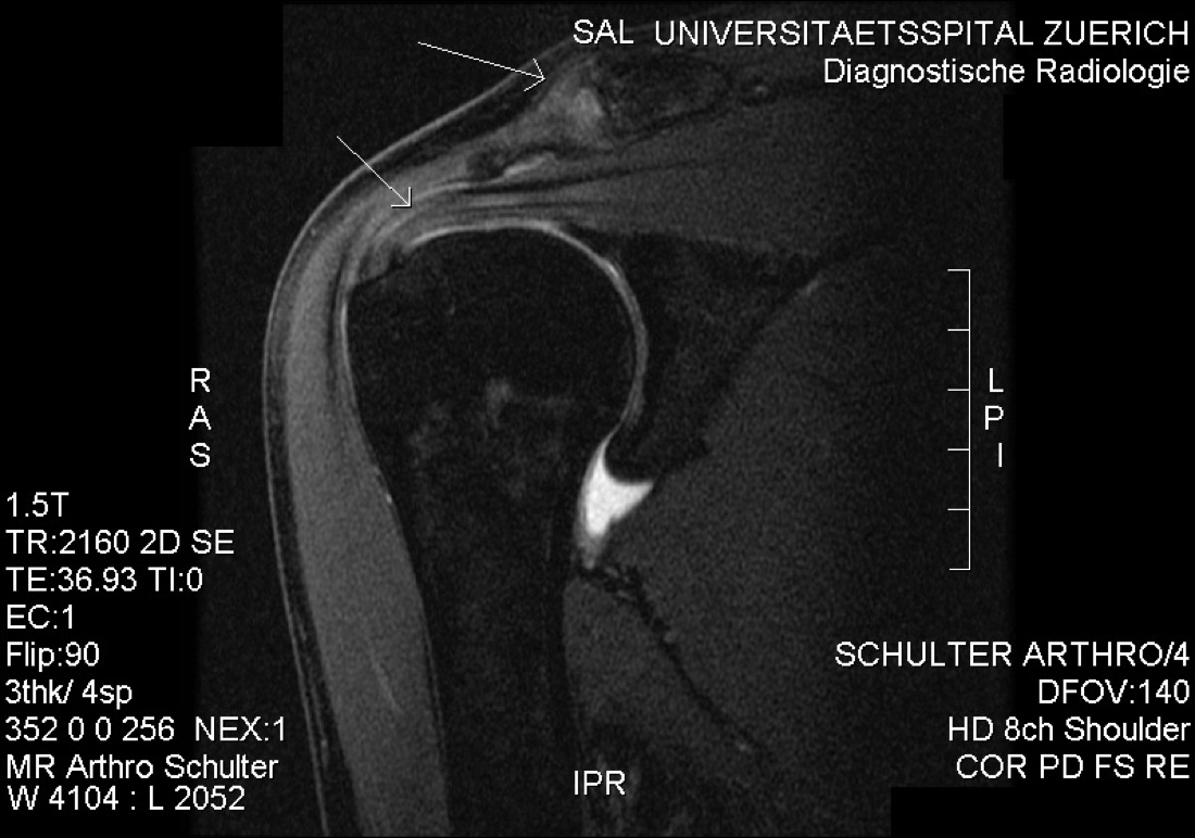
Subacromial impingement syndrome is compression and irritation of the structures within the subacromial space — particularly the supraspinatus tendon and the subacromial bursa — between the undersurface of the acromion and the humeral head. It causes pain, restricted movement and loss of strength, especially with overhead activity. It is most common in patients over 40, in people who work overhead (painters, carpenters) and in athletes who perform repetitive overhead movements (swimmers, tennis players, volleyball players, baseball players). If untreated it may progress to partial rotator cuff tears and later to full-thickness tears.
Anatomy of the Shoulder and the Subacromial Space
The shoulder is a complex region formed by the head of the upper arm bone (humeral head), the shallow socket on the shoulder blade (glenoid), the roof of bone above it (acromion) and the collar bone (clavicle). The narrow space between the acromion and the humeral head is called the subacromial space.
This space contains the supraspinatus tendon (the uppermost tendon of the rotator cuff), the subacromial bursa (a lubricating fluid sac), the long head of biceps tendon and the coracoacromial ligament. In a healthy shoulder these structures move freely; narrowing of the space (impingement) or thickening of the soft tissues (bursitis) causes compression.
Structural (Bony) Causes
- Acromial shape (Bigliani classification): Type I (flat), Type II (curved), Type III (hooked) — Type III is the most prone to impingement
- Acromial osteophyte (bone spur): typically develops on the anterior undersurface
- Osteophytes or arthritis beneath the acromioclavicular joint
- Os acromiale: incomplete fusion of the acromion during development, which increases the risk of impingement
Functional Causes
- Rotator cuff weakness: the humeral head rides upwards and the subacromial space narrows
- Scapular dyskinesis: abnormal positioning of the shoulder blade increases impingement
- Posterior capsular tightness: pushes the humeral head forwards and upwards, causing impingement
- Poor posture: rounded shoulders and a forward head position
Soft-tissue Causes
- Subacromial bursitis: thickening and inflammation of the bursa
- Supraspinatus tendinopathy / tendinitis: degenerative change or inflammatory thickening within the tendon
- Calcific tendinitis: deposition of calcium crystals within the tendon (severe pain during acute flare-ups)
- Partial rotator cuff tear: impingement and tearing create a self-perpetuating cycle
Neer Staging
- Stage I: oedema and haemorrhage (younger patients, reversible) — responds well to non-operative treatment
- Stage II: fibrosis and tendinitis (ages 25–40)
- Stage III: bony change and tendon tearing (over 40) — surgery is often required
Symptoms
- Pain over the outer upper shoulder: worse at night and when lying on the affected shoulder
- Pain with overhead activity: combing hair, hanging washing, taking items from a shelf
- Painful arc: pain between 60° and 120° when raising the arm to the side
- Sensation of impingement: catching or locking during movement
- Loss of strength: particularly with elevation and external rotation
- Crepitus: crackling or catching during movement
- Disturbed sleep: night pain is the last symptom to settle
- Decline in sport-specific performance: with throwing, serving and overhead movements
Diagnosis
Diagnosis is based on a detailed history, physical examination and imaging. The patient's occupation (particularly overhead work), sporting activity, duration and character of pain, night pain, previous shoulder injuries and comorbidities (diabetes, rheumatological disease, smoking) are reviewed.
Examination assesses active and passive range of motion, painful arc, impingement tests (Neer, Hawkins–Kennedy), rotator cuff strength tests (Jobe/empty can for supraspinatus; external rotation for infraspinatus; lift-off for subscapularis), acromioclavicular joint tenderness, scapular dyskinesis and neurovascular status. A subacromial lidocaine injection test may be used to confirm the diagnosis.
Plain radiographs are the first-line investigation; anteroposterior, supraspinatus outlet and axillary views are standard. Ultrasound is a practical, dynamic method for assessing the rotator cuff and subacromial bursa. MRI is the gold standard for assessing supraspinatus tendinopathy, partial or full-thickness tears, subacromial bursitis, acromioclavicular joint arthritis and associated glenohumeral pathology.
Non-operative (Conservative) Treatment
The great majority of patients (around 60%) respond to non-operative treatment. Typical candidates are those with recent or short-lived symptoms, those without a full-thickness rotator cuff tear, and patients at high surgical risk.
- Activity modification: avoiding overhead activity, adjusting work practices
- Non-steroidal anti-inflammatory drugs (NSAIDs)
- Cold therapy / ice
- Physiotherapy: rotator cuff strengthening, scapular stabilisation, posterior capsule stretching, posture correction — this is the most important element
- Subacromial corticosteroid injection: both diagnostic and therapeutic; repeated at most 2–3 times
- PRP (platelet-rich plasma) injection — in selected cases
- For calcific tendinitis: ultrasound-guided aspiration / needle fenestration
- Posture and ergonomic adjustment
Surgical Treatment
Surgery is planned for patients who have not responded to at least 3–6 months of non-operative treatment, whose symptoms significantly affect quality of life, or in whom associated pathology is identified (a prominent osteophyte, acromioclavicular arthritis, a partial cuff tear).
The basis of surgical treatment is arthroscopic subacromial decompression (ASD) and acromioplasty (the Neer procedure). Concomitant procedures (bursectomy, distal clavicle excision, removal of calcific deposits, rotator cuff repair) are added where required.
Advantages
The principal advantage is widening of the subacromial space and permanent relief of compression on the tendons. Chronic pain that has not responded to non-operative treatment improves, functional range of motion is regained, and the risk of progression to a rotator cuff tear is reduced.
- The arthroscopic technique is minimally invasive: small incisions (3–4 portals, each about 5 mm), less pain, lower infection risk and faster recovery
- Diagnosis under direct vision: arthroscopy allows partial rotator cuff tears and glenohumeral pathology not visible on MRI to be seen and treated in the same sitting
- Early motion: passive range-of-motion exercises usually begin the day after surgery, reducing joint stiffness
- Operative time: 30–60 minutes for arthroscopic subacromial decompression; 60–90 minutes with concomitant procedures
- Most patients are treated as day cases
- Less scarring, less pain and faster recovery than open acromioplasty
Disadvantages
- There are surgery-specific risks: wound infection, deep infection, nerve injury (particularly the axillary nerve) and rare anaesthetic complications
- A sling is recommended for 4–6 weeks; active use of the arm is limited during this period
- Frozen shoulder (adhesive capsulitis): may develop despite or after surgery; it is common in patients with diabetes
- Heterotopic ossification (bone formation in soft tissue; rare)
- Inadequate decompression: symptoms may persist if insufficient bone is removed
- Excessive decompression: removing too much of the acromion risks weakness at the deltoid attachment or anterosuperior escape
Why Surgery?
If subacromial impingement syndrome is left untreated, permanent degeneration of the rotator cuff tendons, partial tears and progression to full-thickness tears follow. Once a full-thickness tear develops the tendon retracts and muscle atrophy and fatty infiltration set in, making tendon reconstruction more difficult and the results poorer.
Early surgical decompression eliminates the impingement, improves the healing environment of the tendon and prevents progression of tendon damage in the long term. Timely surgery is preferred in patients who do not respond to non-operative treatment.
Who is a Candidate?
Surgery is offered to patients who have not responded to at least 3–6 months of non-operative treatment (physiotherapy, NSAIDs and at least one to two subacromial injections).
- People in overhead occupations (painters, carpenters) with night pain and functional limitation
- Overhead athletes (swimmers, tennis players, baseball players, volleyball players)
- Patients in whom MRI shows a structural abnormality (Type III acromion, prominent osteophyte, acromioclavicular osteophyte, partial rotator cuff tear, calcific tendinitis)
- Those with a temporary but marked response to subacromial corticosteroid injection whose symptoms recur
- Surgery is not preferred in younger patients before functional causes (scapular dyskinesis, posture) have been corrected
Expected Outcomes
With appropriate indications and compliant rehabilitation, the results of arthroscopic subacromial decompression are good. Between 85% and 90% of patients report a marked reduction in pain and functional improvement. Most return to daily activity within 6–12 weeks and to sport within 4–6 months. Recovery is longer where a rotator cuff repair is also performed (6–12 months).
Factors that adversely affect outcome include smoking, diabetes, advanced age, an associated large rotator cuff tear, previous failed surgery and coexisting frozen shoulder.
Surgery & First 3 Weeks
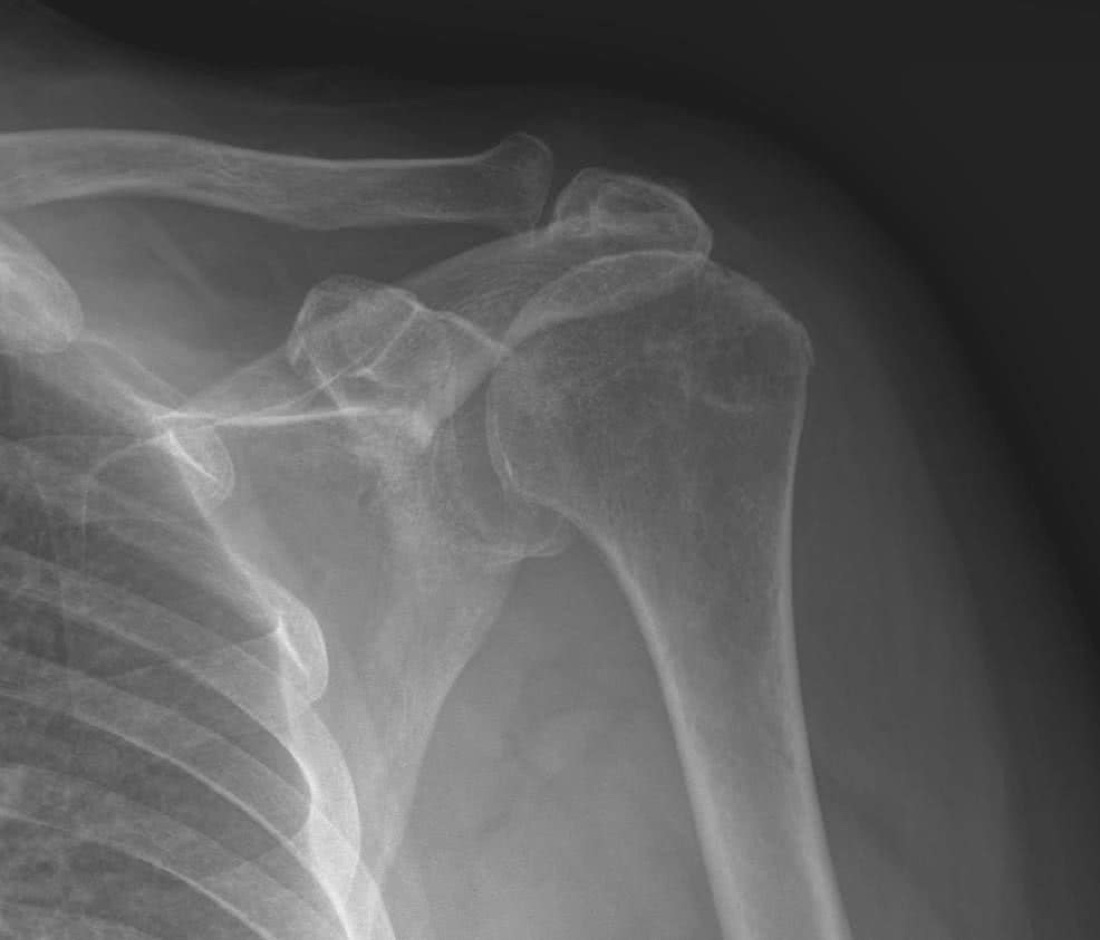
There are two main approaches to the treatment of subacromial impingement syndrome: non-operative (physiotherapy, NSAIDs, injection) and surgical (arthroscopic subacromial decompression — ASD). The choice depends on the duration of symptoms, the severity of pain, functional limitation and associated pathology.
Non-operative Treatment
This is always the first step in subacromial impingement syndrome.
- Activity modification: avoiding overhead movements that trigger pain; ergonomic adjustment at work
- NSAIDs and, if required, a short course of analgesia
- Ice (15–20 minutes, three to four times a day)
- A structured physiotherapy programme (6–12 weeks): posterior capsule stretching, rotator cuff strengthening, scapular stabilisation and posture correction — one of the mainstays of success
- Subacromial corticosteroid injection (at most 2–3 times, with at least 3 months between injections)
- PRP injection in selected cases of tendinitis
- For calcific tendinitis: ultrasound-guided aspiration and lavage
- Posture and ergonomic adjustment (particularly for desk-based workers)
Surgical Treatment — General Information
Surgery is performed under general anaesthesia, frequently combined with an interscalene block. The block is effective for pain control in the first 12–24 hours. Most patients are treated as day cases. Radiographs and MRI are reviewed for preoperative planning, and prophylactic antibiotics are given.
- Duration: 30–60 minutes for isolated ASD; 60–120 minutes with concomitant procedures (bursectomy, distal clavicle excision, removal of calcific deposits, rotator cuff repair)
- Anaesthesia: general anaesthesia plus interscalene block
- Hospital stay: most cases are day cases; an overnight stay may be needed where a cuff repair is performed
Positioning and Surgical Approach
The patient is placed in the beach-chair or lateral decubitus position. In the lateral position the arm is suspended in a traction sling. After preparation and draping, three to four small portals (each 5–6 mm) are made:
- Posterior portal: the viewing (camera) portal, entered between the humeral head and the acromion
- Anterolateral / lateral portal: the working portal, for introducing the shaver and instruments into the subacromial space
- Anterior portal: an additional viewing or working portal
- In some cases additional portals such as the Wilmington or a posterior acromial portal are added
1. Diagnostic Glenohumeral Arthroscopy
- First step: the arthroscope is introduced into the glenohumeral joint through the posterior portal
- The intra-articular structures are examined systematically: labrum, long head of biceps tendon, cartilage surfaces and the undersurface of the rotator cuff (particularly for partial articular-sided supraspinatus tears)
- Associated pathology (SLAP lesion, cartilage damage, biceps tendinitis) is identified
2. Entering the Subacromial Space and Bursectomy
- The arthroscope is directed into the subacromial space
- The hypertrophic subacromial bursa (bursitis) is removed with a shaver; this step is essential for clear visualisation of the subacromial space
- Bursectomy alone can provide marked relief in some patients
3. Acromioplasty (Neer Procedure)
- Osteophytes and the curved or hooked portion of the undersurface of the acromion are removed with a high-speed burr
- Aim: to convert a Type III (hooked) or Type II (curved) acromion into a Type I (flat) acromion
- The undersurface of the coracoacromial ligament is generally preserved or minimally released; complete division may lead to anterosuperior instability
- Modern trend: conservative (minimal) acromioplasty, avoiding excessive bone removal in order to protect the deltoid attachment
4. Concomitant Procedures (by Indication)
- Distal clavicle excision (Mumford procedure): where there is associated acromioclavicular arthritis, 5–8 mm of the distal clavicle is resected
- Removal of calcific deposits: where calcific tendinitis is present, calcium deposits are cleared from the tendon; microfracture or tendon repair may then be added
- Rotator cuff repair: if a partial or full-thickness tear is found, it is repaired with suture anchors in the same sitting
- Biceps tendon procedure (tenotomy or tenodesis): where there is pathology of the long head of biceps
- Labral repair (SLAP repair): where a SLAP lesion is present
- Fixation or partial excision of an os acromiale: if it is contributing to impingement
Implants
No implants are used in isolated subacromial decompression. The following may be used in concomitant procedures:
- Suture anchors (absorbable or titanium; for rotator cuff and labral repair)
- Biceps tenodesis screws or suture anchors (for biceps tenodesis)
- Cannulated screws for os acromiale fixation (a rare indication)
Discharge Information
Most patients are discharged the same day; an overnight stay may be required where a rotator cuff repair or complex procedure has been performed. After surgery the shoulder is supported in a sling. After isolated subacromial decompression the sling is used for a few days to two weeks, whereas 4–6 weeks of full-time use is required if a rotator cuff repair has been added.
Before discharge the physiotherapist teaches how to put on and remove the sling, pendulum (Codman) exercises, elbow, wrist and finger range-of-motion exercises, pain management and transfers. After isolated ASD, passive and active-assisted shoulder range-of-motion exercises usually begin within the first 1–7 days; the protocol is more restricted if a cuff repair has been added.
Ice for 15–20 minutes three to four times a day is recommended after discharge. Analgesics, anti-inflammatories and gastric protection are prescribed. Routine anticoagulation is not given for shoulder surgery. Dressings are changed regularly; it is important not to wet the wound for the first 10–14 days, and sutures are removed on day 10–14. Sleeping semi-upright reduces pain in the first weeks.
Early Postoperative Period (First 3 Weeks)
The aims during this period are wound healing, control of swelling, pain management, preserving elbow, wrist and finger movement, minimising muscle atrophy and preventing shoulder stiffness (frozen shoulder). Weekly review is recommended.
- Wound monitoring: discharge, marked redness, increased warmth or wound edge separation are monitored. It is important not to wet the wound for the first 10–14 days.
- Sling use: a few days to two weeks after isolated ASD; 4–6 weeks full time (including at night) if a rotator cuff repair has been added. Active elevation of the arm is prohibited in the early weeks.
- Early passive movement: to prevent frozen shoulder, passive pendulum (Codman) and gentle passive flexion and external rotation exercises begin within the first 1–7 days with the surgeon's permission; repeat three to four times a day.
- Elbow, wrist and finger movement: performed three to four times a day within the sling; this reduces swelling and prevents stiffness in the adjacent joints.
- Pain: once the interscalene block wears off, prescribed medication, ice and a semi-upright sleeping position control the pain. Night pain is the last symptom to settle.
- Nerve monitoring: the axillary nerve is at risk during shoulder surgery; sensation over the outer upper arm and deltoid contraction are checked.
Follow-up
- Days 10–14: suture removal
- Weeks 4–6: progression of movement and activity
- Month 3: functional review
- Month 6 and, if required, year 1
Risks & Complications
Arthroscopic subacromial decompression is one of the safest procedures in modern shoulder surgery; the rate of serious complications is below 1%. Nevertheless, every operation carries specific risks.
Risks Specific to Non-operative Treatment
- NSAID side effects: gastric bleeding, impaired kidney or liver function, cardiovascular risk
- Repeated subacromial corticosteroid injection: tendon weakening, local skin changes, raised blood glucose (in patients with diabetes), infection (very rare)
- Delayed diagnosis or treatment: progression of a partial rotator cuff tear to a full-thickness tear
- Development of frozen shoulder (adhesive capsulitis): restricted movement and prolonged pain
- Prolonged reduction in quality of life
Risks Specific to Surgery — Common (Under 5%)
- Superficial wound infection (usually treated with antibiotics)
- Postoperative pain, swelling and temporary bruising
- Temporary numbness or tenderness at the portal sites
- Transient symptoms related to anaesthesia such as nausea and dizziness
- Temporary numbness after an interscalene block; very rarely, diaphragmatic paralysis
- Temporary shoulder stiffness — resolves with regular physiotherapy
Risks Specific to Surgery — Less Common
- Deep infection: 0.5–2%; may require revision, irrigation and prolonged antibiotics; the most common organism is Cutibacterium acnes
- Nerve injury: the axillary nerve is the nerve most at risk in shoulder surgery; numbness over the outer upper arm or deltoid weakness may develop
- Temporary or permanent injury to elements of the brachial plexus (particularly from traction in the lateral position)
- Vascular injury (very rare)
- Fluid extravasation: leakage of high-pressure fluid into the subacromial space during arthroscopy, causing soft-tissue swelling and, rarely, airway compression
- Deep vein thrombosis (DVT) — very rare in the upper limb
Complications Specific to the Procedure
- Inadequate decompression: persistent symptoms because insufficient bone was removed; revision may be required
- Excessive decompression: weakness at the deltoid attachment from removing too much of the acromion
- Anterosuperior escape: superior migration of the humeral head due to insufficiency of the acromion and coracoacromial ligament (particularly where there is an associated cuff tear)
- Acromial stress fracture (very rare)
- Frozen shoulder (adhesive capsulitis): develops in 5–10% of patients after surgery, particularly in those with diabetes. Treatment: intensive physiotherapy, intra-articular injection and, in advanced cases, manipulation or arthroscopic capsular release
- Heterotopic ossification: bone formation in soft tissue, which may restrict movement; rare
Persistent or Recurrent Symptoms
- Incorrect diagnosis: surgery will fail if the true cause is scapular dyskinesis, cervical radiculopathy or another problem
- Failure to treat associated pathology (for example a missed rotator cuff tear)
- Excessive traumatic or occupational loading (inadequate activity modification)
Factors that Increase Risk
- Smoking (increases the risk of wound healing problems, frozen shoulder and infection)
- Poorly controlled diabetes (risk of frozen shoulder and infection)
- Immunosuppressive therapy and rheumatological disease
- Advanced age; previous shoulder surgery
- Coexisting cervical (neck) problems
- Active or recently treated infection (dental, urinary, skin)
- Non-compliance with the rehabilitation programme
Contact Your Doctor Immediately If You Have
- Sudden increase in pain or swelling
- Fever above 38 °C
- Discharge, malodour or separation of the wound
- Numbness, tingling or loss of strength in the arm, shoulder or hand
- Bruising, coldness or pallor of the hands or fingers
- Shortness of breath or chest pain
- Marked swelling of the neck or face (suspected fluid extravasation)
Long-term Outcomes
With appropriate indications and compliant rehabilitation, the results of arthroscopic subacromial decompression are good. Between 75% and 90% of patients report a marked reduction in pain and functional improvement, and patient satisfaction is high at 5–10 year follow-up.
Factors that adversely affect outcome include coexisting cervical pathology or scapular dyskinesis, an associated large rotator cuff tear, smoking, diabetes, advanced age and non-compliance with rehabilitation. Where a rotator cuff repair has also been performed, the long-term result depends largely on healing of the cuff. Postoperative physiotherapy, posture and occupational ergonomics are among the mainstays of success.
Rehabilitation
Rehabilitation after subacromial decompression is the most important part of recovery. The aims are to control pain, preserve and restore shoulder range of motion, strengthen the rotator cuff and scapular stabilisers, correct posture, and return the patient to daily life and sporting activity. Because frozen shoulder is one of the most important complications of shoulder surgery, starting passive range-of-motion exercises as soon as it is safe to do so is critical.
Non-operative Treatment (Without Surgery)
- Weeks 0–2: Activity modification (avoiding overhead movements), NSAIDs, ice. Pendulum (Codman) exercises and gentle passive range-of-motion work. Posterior capsule stretching.
- Weeks 2–6: Structured physiotherapy: active-assisted and active range of motion, isometric rotator cuff exercises, scapular stabilisation, posture correction.
- Weeks 6–12: Resisted rotator cuff and scapular strengthening with a resistance band. Isokinetic exercise.
- Months 3–6: Sport-specific work; graduated return to overhead activity. Ergonomic adjustment is maintained. If symptoms persist, surgical treatment is considered.
After Isolated Subacromial Decompression (ASD)
- Week 0–1: Sling, ice and elevation. Active elbow, wrist and finger range-of-motion exercises. With the surgeon's permission, pendulum and gentle passive flexion and external rotation exercises are started.
- Weeks 1–3: The sling is discontinued gradually (as tolerated during the day). Passive and active-assisted shoulder range-of-motion work intensifies. Scapular isometric exercises begin. Full passive range of motion is the target.
- Weeks 3–6: Active range-of-motion exercises begin; full active range is the target. Light isometric rotator cuff exercises. Postural work.
- Weeks 6–12: Resisted rotator cuff and scapular strengthening with a resistance band. Light lifting exercises. Cycling and swimming (once the wound has healed).
- Months 3–6: Advanced strengthening. Graduated return to sport-specific drills and throwing mechanics.
- Months 4–6: Full return to sporting and overhead occupational activity.
Rehabilitation When a Rotator Cuff Repair is Added
If a rotator cuff repair was also performed, the rehabilitation protocol is more restricted. The detailed protocol is individualised according to the size of the tear.
- Weeks 0–4: Sling full time (including at night). Passive range-of-motion exercises only (within the limits set by the surgeon). Active movement and resisted work are prohibited.
- Weeks 4–6: The sling is discontinued. Exercises against gravity begin.
- Weeks 6–12: Active-assisted and active range-of-motion exercises begin. Scapular isometric exercises.
- Months 3–4: Light strengthening (resistance band). Weight lifting is prohibited.
- Months 4–6: Advanced strengthening; full return to daily activity.
- Months 6–12: Full return to sport and overhead activity with the surgeon's approval.
Criteria for Return to Sport and Activity
- Full, pain-free shoulder range of motion (flexion ≥150°, abduction ≥150°, external rotation ≥60°)
- Rotator cuff and scapular muscle strength at least 85–90% of the opposite shoulder
- No swelling or pain after activity
- Pain-free completion of sport-specific drills
- Control of throwing mechanics
- Approval of the surgeon and physiotherapist
Recommended Activities
- Stationary cycling, elliptical trainer, walking
- Swimming (once the wound has healed, progressively)
- Low-impact conditioning and strengthening exercise
- Pilates and yoga (with the surgeon's permission; overhead poses may be limited initially)
- Light recreational activity
Activities to Avoid in the First 3–6 Months
- Lifting weights overhead
- Sudden pulling or tugging movements
- Throwing sports (baseball, volleyball, handball)
- Tennis (serving and smashing)
- Contact sport
- Lying on the operated shoulder (particularly for the first 3 months)
- Prolonged sitting in a poor posture and repetitive overhead movements at work
Lifestyle and Recovery Advice
- Perform your home exercise programme every day — passive range-of-motion and postural exercises in particular are the most effective way to prevent frozen shoulder
- Do not smoke (risk of wound healing problems and frozen shoulder)
- Maintain tight blood glucose control if you have diabetes (risk of frozen shoulder)
- Posture and ergonomics: pay attention to monitor height and seating posture when working at a desk
- Occupational adjustment: minimise repetitive overhead movements; seek workplace ergonomic advice if necessary
- Sleeping semi-upright (supported with pillows) reduces pain in the first weeks
- Avoid lying on the operated shoulder for the first 3 months
- Attend your follow-up appointments (weeks 4–6, month 3, month 6)
Expected Outcomes
Most patients return to pain-free daily activity 6–12 weeks after subacromial decompression. Night pain is one of the first symptoms to improve; most patients regain undisturbed sleep within the first weeks.
Full return to sporting and overhead occupational activity occurs at 3–4 months. If a rotator cuff repair was also performed, recovery extends to 6–9 months. Patient satisfaction is high with the modern arthroscopic technique, appropriate indications and disciplined rehabilitation. Posture correction, physiotherapy and occupational ergonomics are critical to maintaining the result long term.
Is This Treatment Right for You?
Every patient's condition is different. After a detailed examination and imaging, the treatment plan that suits you best will be prepared.
Would you like more information about Subacromial Impingement Syndrome?
Book Appointment